Unsupported Browser
The American College of Surgeons website is not compatible with Internet Explorer 11, IE 11. For the best experience please update your browser.
Menu
Become a member and receive career-enhancing benefits

Our top priority is providing value to members. Your Member Services team is here to ensure you maximize your ACS member benefits, participate in College activities, and engage with your ACS colleagues. It's all here.

Become a Member
Become a member and receive career-enhancing benefits

Our top priority is providing value to members. Your Member Services team is here to ensure you maximize your ACS member benefits, participate in College activities, and engage with your ACS colleagues. It's all here.

Membership Benefits
ACS
Case Reviews in Surgery

A Hemodynamically Stable Patient with a Transmediastinal Gunshot Wound

A Hemodynamically Stable Patient with a Transmediastinal Gunshot Wound

March 9, 2020

Abstract

Background

A 29-year old man was evaluated after sustaining a gunshot wound to his right axilla. He was hemodynamically stable without respiratory distress. Chest X ray revealed a bullet overlying the left mediastinum with an apparent trajectory concerning for cardiac injury, while FAST exam did not demonstrate pericardial effusion.

Summary

Initial X-ray findings were misleading in this patient, and we were able to rule out clinically significant mediastinal injuries with cross-sectional imaging, endoscopy, bronchoscopy, and a pericardial window. It appears that the bullet traversed both pleural spaces and his mediastinum anterior to the great vessels, causing a pneumothorax that allowed the bullet to travel freely in his pleural space.

Conclusion

The hemodynamically stable patient with a transmediastinal gunshot wound presents a clinical challenge, and isolated imaging findings can be misleading. A straight line between a gunshot wound and bullet on X ray does not always determine trajectory. When an urgent surgical intervention is not indicated, less invasive options to evaluate the hemodynamically stable patient with transmediastinal gunshot wound include CTA, bronchoscopy, upper endoscopy, esophagram, echocardiogram, VATS, pleuroscopy, and pericardial window.

Key Words

transmediastinal gunshot wound; pleural space foreign body; lung injury; pericardial window


Case Description

A 29-year old man was transferred to our level 1 trauma center from a community emergency department after sustaining a gunshot wound to his right axilla (marked by a closed paperclip signifying an anterior wound on chest X ray in Fig 1). He was found to have a contralateral, left-sided pneumothorax on chest X ray at the community emergency department and a left-sided tube thoracostomy was inserted prior to transfer. On arrival at our trauma center, the patient was hemodynamically normal and neurologically intact with minimal hypoxia and no respiratory distress. Trauma survey was notable only for a single gunshot wound to the right axilla along the anterior axillary line. Focused assessment with sonography in trauma (FAST) exam was unremarkable without evidence of pericardial effusion. Chest X ray revealed a right-sided pulmonary contusion, as well as a bullet overlying the left mediastinum. The proposed trajectory from entry to bullet is Transmediastinal, as indicated by the dashed line in Figure 1.

Figure 1: Chest X ray of patient with Transmediastinal gunshot wound. Dotted red line depicts bullet trajectory implied by skin wound (marked by paper clip) and location of bullet on X ray.

Figure 1: Chest X ray of patient with Transmediastinal gunshot wound. Dotted red line depicts bullet trajectory implied by skin wound (marked by paper clip) and location of bullet on X ray.

Discussion

In truncal penetrating injury, the bullet trajectory (wound-to-bullet) usually predicts injury pattern. In this patient, a straight line from the skin wound to the bullet's location on X ray raised concern for cardiac, major vascular, aerodigestive and/or thoracic vertebral injuries. The patient's hemodynamic stability, neurologically intact examination and lack of pericardial effusion on FAST or significant right-sided hemothorax on chest X ray were reassuring but did not rule out life-threatening injury.

There is little medical literature to guide management of the hemodynamically stable patient with transmediastinal penetrating trauma. Several case series have been published supporting institutional protocols to evaluate patients nonoperatively using chest X ray, chest CT and FAST exam or transthoracic echocardiogram, followed by angiogram, esophagram and esophagoscopy, bronchoscopy and pericardial window as indicated by initial study results.1-3 Nagy et al. attempted to differentiate anterior mediastinal injuries that could be evaluated with echocardiogram and possible pericardial window from posterior mediastinal injuries that required angiogram, esophagram and esophagoscopy with possible bronchoscopy.1 A subsequent Western Trauma Association Critical Decisions in Trauma algorithm for patients with penetrating chest trauma recommends evaluation of hemodynamically stable patients with transmediastinal penetrating trauma using CTA and possible bronchoscopy, upper endoscopy and esophagram, and recommends evaluation of suspected cardiac injuries with FAST exam, followed by VATS, pleuroscopy, or pericardial window if the diagnosis remains unclear, especially in the context of a hemothorax.4

Given his clinical stability, we opted to send our patient for chest CT with intravenous contrast, which revealed a comminuted right second rib fracture, right upper lung contusion with trace right hemopneumothorax, anterior mediastinal hematoma, and left upper lung contusion with left hemothorax. There was no evidence of great vessel injury, active extravasation or pericardial effusion. The bullet was located much more inferiorly, in the left diaphragmatic sulcus at the left lung base. We believe that the bullet traversed both lungs and the superior anterior mediastinum (see red lines in Figure 2 and Figure 3), striking the left second rib and coming to rest in the pleural space along this trajectory. From there, the bullet apparently moved dependently within the pleural space and came to rest in the posterior sulcus.

Figure 2. Coronal CT image of patient with Transmediastinal gunshot wound. Solid red line depicts bullet trajectory based on tissue injury on CT scan.

Figure 2. Coronal CT image of patient with Transmediastinal gunshot wound. Solid red line depicts bullet trajectory based on tissue injury on CT scan.

Figure 3. Axial CT image of patient with Transmediastinal gunshot wound depicting the bullet trajectory. Solid red line depicts bullet trajectory based on tissue injury on CT scan.

Figure 3. Axial CT image of patient with Transmediastinal gunshot wound depicting the bullet trajectory. Solid red line depicts bullet trajectory based on tissue injury on CT scan.

Bullet migration is a rare but well-documented phenomenon, with published case reports of bullets traveling through blood vessels,5 the gastrointestinal tract,6 the pericardium,7 and the dural space.8 Accurately identifying and appropriately managing these injuries requires a high index of suspicion, close attention to atypical symptoms such as perfusion abnormalities and neurologic deficits and use of multiple, complementary imaging modalities.

The patient was taken urgently to the operating room for further evaluation, as the CT findings did not guarantee a linear bullet trajectory across the anterior mediastinum superior to the heart and did not definitively rule out cardiac, esophageal, or tracheobronchial injuries. Despite his negative FAST exam, the patient's bilateral hemothoraces raised the possibility of an occult cardiac injury decompressing into his pleural spaces (Figure 4).

Figure 4. Axial CT image of patient with Transmediastinal gunshot wound depicting bullet location. Solid yellow line indicates the position of the bullet in the left diaphragmatic sulcus, posterior to the left kidney on CT scan.

Figure 4. Axial CT image of patient with Transmediastinal gunshot wound depicting bullet location. Solid yellow line indicates the position of the bullet in the left diaphragmatic sulcus, posterior to the left kidney on CT scan.

The patient's malpositioned left tube thoracostomy was replaced and we opted to place a right-sided tube thoracostomy with concern for hemothorax accumulation or occult pneumothorax that could worsen with positive pressure ventilation. An Eastern Association for the Surgery of Trauma Practice Management Guideline cites level III evidence to suggest that occult pneumothoraces may be observed in stable patients despite positive pressure ventilation,9 which has been supported by a randomized controlled trial conducted by the Canadian Trauma Trials Collaborative.10 However, we were not convinced that those findings applied to this patient with a penetrating lung injury and suspected cardiac injury.

Next we performed pericardial window via a subxyphoid approach to rule out cardiac injury, which was negative. Subsequent upper endoscopy and bronchoscopy did not reveal esophageal or tracheobronchial injury, respectively. Post-operatively, the patient remained intubated and was admitted to the ICU for ventilator support of his hypoxemic respiratory failure in the context of bilateral pulmonary contusions. He had an uncomplicated hospital course, was extubated on post-injury day 3 and was discharged home on post-injury day,9

A final follow-up chest X ray (Figure 5), obtained three weeks after the injury, shows resolution of the lung injury with the bullet still positioned in the left diaphragmatic sulcus. We did consider performing a video-assisted thoracoscopic bullet retrieval, as has been previously described,11 but opted not to as the bullet was not causing ongoing pain or inflammation.

Figure 5. Follow-up chest X ray of patient with Transmediastinal gunshot wound. Solid yellow line indicates the position of the bullet on X ray.

Figure 5. Follow-up chest X ray of patient with Transmediastinal gunshot wound. Solid yellow line indicates the position of the bullet on X ray.

Conclusion

In this case of a hemodynamically stable patient with a transmediastinal gunshot wound, given the patient's clinical stability evaluation using CT, pericardial window, endoscopy and bronchoscopy allowed us to rule out clinically significant mediastinal injury without a more morbid surgical exploration.

Lessons Learned

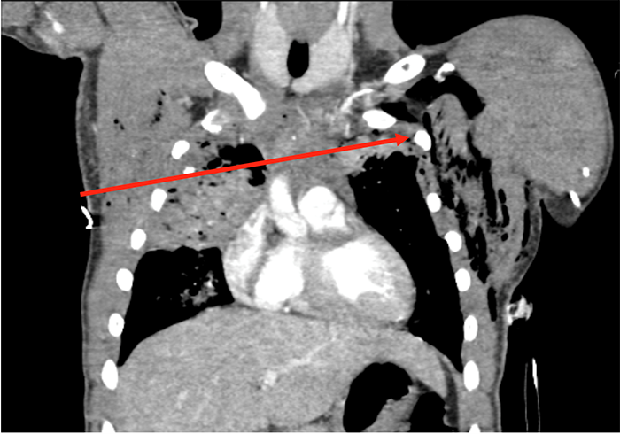
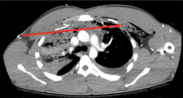
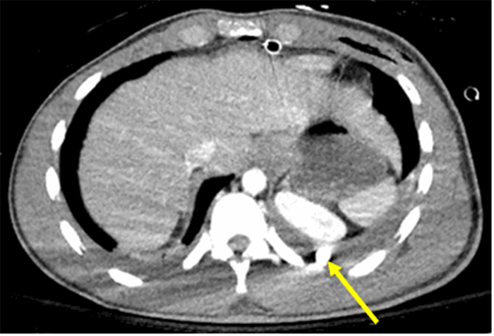
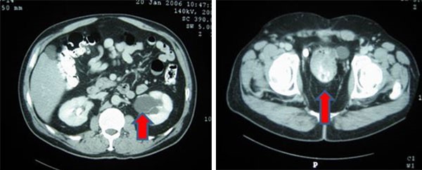
While implied trajectory on X rays can suggest injury patterns and guide further investigation, it is important to remember that bullets do not always follow linear paths. In particular, bullets can migrate freely in the pleural space in the context of a hemopneumothorax.

Authors

Adam D. Laytin, MD, MPH
Division of Traumatology, Surgical Critical Care & Emergency Surgery
Department of Surgery
University of Pennsylvania; Philadelphia, PA

Michael A. Vella, MD, MBA
Division of Traumatology, Surgical Critical Care & Emergency Surgery
Department of Surgery
University of Pennsylvania; Philadelphia, PA

Jose L. Pascual, MD, PhD, FACS
Division of Traumatology, Surgical Critical Care & Emergency Surgery
Department of Surgery
University of Pennsylvania; Philadelphia, PA

Niels D. Martin, MD, FACS
Division of Traumatology, Surgical Critical Care & Emergency Surgery
Department of Surgery
University of Pennsylvania; Philadelphia, PA

Correspondence Author

Niels D. Martin, MD, FACS, FCCM
Division of Traumatology, Surgical Critical Care & Emergency Surgery
Perelman School of Medicine at the University of Pennsylvania
51 North 39th St, Medical Office Building Suite 120
Philadelphia, PA 19104
Phone: 215-662-7323
Email: Niels.Martin@uphs.upenn.edu

Disclosure Statement

Conflicts of Interest and Source of Funding: No author has a conflict of interest, competing interest or financial disclosure to declare. All authors have met criteria for authorship and ethical adherence. There were no sources of funding for this project.

References

  1. Nagy KK, Roberts RR, Smith RF, Joseph KT, An GC, Bokhari F, Barrett J. Transmediastinal gunshot wounds: are "stable" patients really stable? World J Surg. 2002;26(10):1247-50.
  2. Stassen NA, Lukan JK, Spain DA, Miller FB, Carrillo EH, Richardson JD, Battistella FD. Reevaluation of diagnostic procedures for transmediastinal gunshot wounds.J Trauma. 2002;53(4):635-8; discussion 8.
  3. Ibirogba S, Nicol AJ, Navsaria PH. Screening helical computed tomographic scanning in haemodynamic stable patients with transmediastinal gunshot wounds. Injury. 2007;38(1):48-52.
  4. Karmy-Jones R, Namias N, Coimbra R, Moore EE, Schreiber M, McIntyre R, Jr., Croce M, Livingston DH, Sperry JL, Malhotra AK, et al. Western Trauma Association critical decisions in trauma: penetrating chest trauma. J Trauma Acute Care Surg. 2014;77(6):994-1002.
  5. Treto K, Bhullar IS, Lube MW. Iliac Artery Bullet Embolus after Isolated Thoracic Ballistic Injury. Am Surg. 2017 Jul 1;83(7):e253-254.
  6. Hughes JJ. Bullet injury to the esophagus detected by intestinal migration. J Trauma. 1987 Dec;27(12):1362-4.
  7. Davis RE, Bruno AD, Larsen WB, Sugimoto JT, Gaines RD. Mobile intrapericardial bullet: case report and review of the literature. J Trauma. 2005 Feb;58(2):378-80.
  8. Todnem N, Hardigan T, Banerjee C, Alleyne CH. Cephalad Migration of Intradural Bullet from Thoracic Spine to Cervical Spine. World Neurosurg. 2018;119:6-9.
  9. Mowery NT, Gunter OL, Collier BR, Diaz JJ, Jr., Haut E, Hildreth A, Holevar M, Mayberry J, Streib E. Practice management guidelines for management of hemothorax and occult pneumothorax. J Trauma. 2011;70(2):510-8.
  10. Kirkpatrick AW, Rizoli S, Ouellet JF, Roberts DJ, Sirois M, Ball CG, Xiao ZJ, Tiruta C, Meade M, Trottier V, et al. Occult pneumothoraces in critical care: a prospective multicenter randomized controlled trial of pleural drainage for mechanically ventilated trauma patients with occult pneumothoraces. J Trauma Acute Care Surg. 2013;74(3):747-54; discussion 54-5.
  11. Williams CG, Haut ER, Ouyang H, Riall TS, Makary M, Efron DT, Cornwell EE. Video-assisted thoracic surgery removal of foreign bodies after penetrating chest trauma. J Am Coll Surg. 2006;202(5):848-52.