Discussion
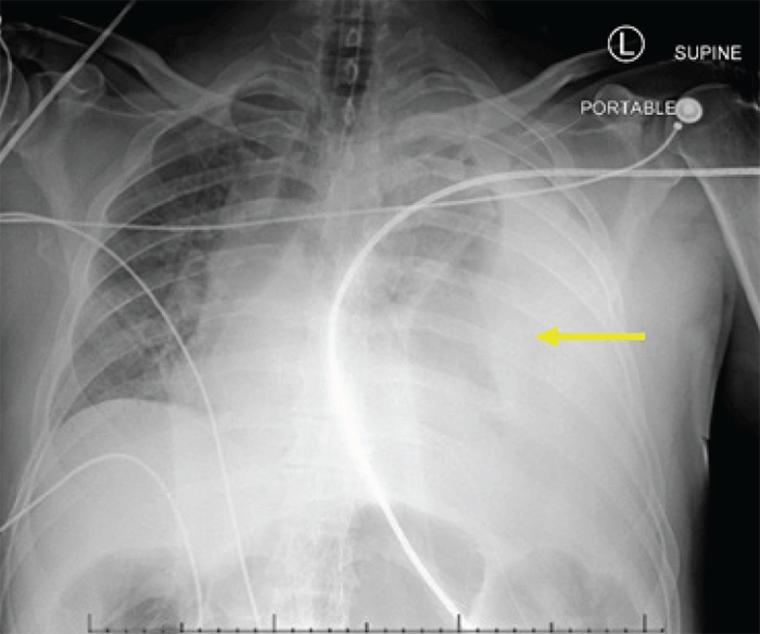
The case describes a trauma patient who experienced two distinct, rare clinical complications: delayed massive hemothorax with tension physiology and paraplegia complicating endovascular aortic repair.
Upon review, there were several opportunities for improved clinical decision-making. First, the standard of care for massive hemothorax is operative control of bleeding and not angioembolization. This conclusion is especially true with any history of aortic stenting, which may prohibit cannulization of the bleeding intercostal artery. Nonetheless, a discussion between the trauma surgery and interventional radiology teams yielded a decision to attempt endovascular control of bleeding before proceeding to the operating room. Although no definite bleeding source was identified on angiography, the chest tube output diminished significantly after adequate resuscitation with blood products, and any operative exploration was ultimately aborted.
Second, massive transfusion protocol should have been initiated earlier. After hemodynamic instability with an identified source of ongoing bleeding, earlier rapid transfusion may have helped maintain perfusion of the spinal cord.
Delayed hemothorax is an underappreciated and relatively common complication after blunt thoracic trauma. In one study of 1,382 patients sustaining any type of chest trauma, radiographically apparent effusions were observed in 11% at 14 days.1 In comparison, pneumonia complicated only 2% of patients sustaining chest trauma. Of the patients with pleural effusions, only 50% complained of chest pain and only 22% of dyspnea. However, only 3% required drainage, equal to 0.36% of the total study population. Age greater than or equal to 70 years conferred the highest relative risk of developing delayed hemothorax, followed by the presence of high or midrib fracture, age 45‒70 years, and presence of at least three rib fractures (specificity 90.7%, sensitivity 33.9%, receiver operator curve 0.78). Another study found that 92% of cases of delayed hemothorax were preceded by some clinically apparent symptom, commonly dyspnea, chest pain, coughing, or diaphoresis.2 The time delay from initial injury to hemothorax can vary significantly; one study found that identification of new pleural effusion occurred anywhere from 18 hours to 11 days after traumatic injury.3 Rib fractures on initial presentation post-thoracic injury should increase suspicion of the possibility of developing a hemothorax later in the course of care. Any number of rib fractures has been associated with a higher risk of delayed bleeding into the pleural space. The cause of bleeding is most commonly laceration of the diaphragm, intercostal artery, and/or phrenic artery.3‒6
Massive hemothorax, generally defined as the evacuation of more than 1500 ml of blood immediately after tube thoracostomy or 200 ml/hr of output for four hours, is an infrequent complication of blunt thoracic trauma. Massive hemothorax requires surgical intervention to identify and stop the source of bleeding.
Delayed massive hemothorax after a blunt thoracic injury is an extremely rare entity. In a single-institution study of 1278 patients sustaining blunt thoracic trauma, only five (0.4%) had delayed massive hemothorax. All underwent emergent thoracotomy and were found to be bleeding from a laceration of the diaphragm by a sharp fragment from a nearby rib fracture.
Any patient with blunt thoracic trauma experiencing new symptoms after their initial presentation should be evaluated with a chest radiograph. Early recognition of and intervention for massive hemothorax cannot be understated due to the risk of significant morbidity or mortality. Untreated, retained hemothorax may lead to complications such as empyema and fibrothorax and more extended hospital stays.1 Therefore, continuous evaluation for hemothorax, especially with potentially delayed presentation, can facilitate its identification and resolution and confer a more favorable prognosis with timely tube thoracostomy and adequate resuscitation.3 If there is aortic damage requiring thoracic endovascular aortic repair (EVAR), the obstructing endovascular stent-grafts may render lacerated intercostal arteries unable to be opacified or embolized via angiography, preventing control of the bleeding.
Traumatic thoracic aortic injury can be immediately life-threatening and can lead to aortic dissection, rupture, or thrombus, and warrants prompt surgical intervention. Compared to open surgical repair, EVAR has been shown to confer significantly lower mortality and risk of delayed postoperative paraplegia, one of the most commonly recognized complications of thoracic aortic aneurysm repair.7 However, the risk of paraplegia after EVAR is not insignificant, with a reported incidence from 0.21 to 15%.7‒11 Cheung et al.12 also found a considerable variation in the timing of delayed-onset paraplegia, with the initial episode occurring 6.4 to 110 hours after surgery and a second episode occurring an average of 176 hours postoperatively. Another study by Maniar et al.10 reported delayed paraplegia occurring up to 27 days after EVAR, with most cases associated with a documented episode of hypotension.
Lower extremity paraparesis and paraplegia result from spinal cord ischemia (SCI). The incidence of SCI is much greater for endovascular repair of the thoracic aorta, which can be as high as 12% compared to the abdominal aorta. SCI has been independently associated with preoperative renal insufficiency.13,14 Other contributing factors to the development of SCI include perioperative hypotension, increased cerebrospinal fluid (CSF) pressure, and occlusion of supplying arteries, such as the artery of Adamkiewicz or intercostal arteries by stent-grafts.10,12
Damage to or occlusion of the artery of Adamkiewicz, a posterior intercostal artery branch, can rarely result in anterior cord syndrome. This artery is the most prominent thoracic radicular artery. It can be found between the T9 and T12 levels in most individuals, supplying the lower, anterior two-thirds of the spinal cord via the anterior spinal artery. This territory supplied by the anterior spinal artery is the most common location of SCI because of its single blood supply, unlike the dual supply from the two posterior spinal arteries that feed the posterior spinal cord. Spinal cord infarction is also most common at the lower thoracic and upper lumbar levels, manifesting as complete lower extremity motor paralysis and loss of temperature and pain perception distal to the lesion, as well as urinary and rectal incontinence or retention. Since the posterior columns are spared, light touch, vibration, and proprioception are preserved.15,16
Patients with delayed-onset paraplegia have been found to have an increased chance of recovery compared to those in whom paraplegia was diagnosed upon emergence from anesthesia, and most patients with delayed-onset paraplegia experience full neurologic recovery.12,14 Despite the possible reversibility of this complication, watchful postoperative monitoring is imperative in all patients who have undergone EVAR. Avoiding even transient hypotension with blood pressure augmentation to higher mean arterial pressures and decreasing CSF pressures have been shown to reduce the risk of paraplegia.10,14 Decreasing CSF pressure using spinal drains has also been demonstrated to protect against spinal cord ischemia during EVAR.17
Conclusion
Delayed massive hemothorax is an extremely rare clinical entity. When it occurred in a patient with a recent endovascular stent for aortic injury, the concomitant hypotension caused infarction of the anterior thoracic spinal cord resulting in paraplegia.
Lessons Learned
Delayed massive hemothorax is a rare but highly morbid complication of blunt thoracic trauma, and any new complaint should be thoroughly investigated with chest radiography. Paraplegia is a rare but highly morbid complication following thoracic endovascular repair, and every effort should be made to avoid postoperative hypotension
References
- Émond M, Guimont C, Chauny JM, et al. Clinical prediction rule for delayed hemothorax after minor thoracic injury: a multicentre derivation and validation study. CMAJ Open. 2017;5(2):E444-E453. doi:10.9778/cmajo.20160096
- Simon BJ, Chu Q, Emhoff TA, Fiallo VM, Lee KF. Delayed hemothorax after blunt thoracic trauma: an uncommon entity with significant morbidity. J Trauma. 1998;45(4):673-676. doi:10.1097/00005373-199810000-00005
- Chang SW, Ryu KM, Ryu JW. Delayed massive hemothorax requiring surgery after blunt thoracic trauma over a 5-year period: complicating rib fracture with sharp edge associated with diaphragm injury. Clin Exp Emerg Med. 2018;5(1):60-65. Published 2018 Mar 30. doi:10.15441/ceem.16.190
- Al-Koudmani I, Darwish B, Al-Kateb K, Taifour Y. Chest trauma experience over eleven-year period at al-mouassat university teaching hospital-Damascus: a retrospective review of 888 cases. J Cardiothorac Surg. 2012;7:35. Published 2012 Apr 19. doi:10.1186/1749-8090-7-35
- Curfman KR, Robitsek RJ, Salzler GG, et al. Massive hemothorax caused by a single intercostal artery bleed ten days after solitary minimally displaced rib fracture. Case Rep Surg. 2015;2015:120140. doi:10.1155/2015/120140
- Racine S, Émond M, Audette-Côté JS, et al. Delayed complications and functional outcome of isolated sternal fracture after emergency department discharge: a prospective, multicentre cohort study. CJEM. 2016;18(5):349-357. doi:10.1017/cem.2016.326
- Xenos ES, Abedi NN, Davenport DL, et al. Meta-analysis of endovascular vs open repair for traumatic descending thoracic aortic rupture. J Vasc Surg. 2008;48(5):1343-1351. doi:10.1016/j.jvs.2008.04.060
- Berg P, Kaufmann D, van Marrewijk CJ, Buth J. Spinal cord ischaemia after stent-graft treatment for infra-renal abdominal aortic aneurysms. Analysis of the Eurostar database. Eur J Vasc Endovasc Surg. 2001;22(4):342-347. doi:10.1053/ejvs.2001.1470
- Buth J, Harris PL, Hobo R, et al. Neurologic complications associated with endovascular repair of thoracic aortic pathology: Incidence and risk factors. a study from the European Collaborators on Stent/Graft Techniques for Aortic Aneurysm Repair (EUROSTAR) registry. J Vasc Surg. 2007;46(6):1103-1111. doi:10.1016/j.jvs.2007.08.020
- Maniar HS, Sundt TM 3rd, Prasad SM, et al. Delayed paraplegia after thoracic and thoracoabdominal aneurysm repair: a continuing risk. Ann Thorac Surg. 2003;75(1):113-120. doi:10.1016/s0003-4975(02)04494-6
- Naughton PA, Park MS, Morasch MD, et al. Emergent repair of acute thoracic aortic catastrophes: a comparative analysis. Arch Surg. 2012;147(3):243-249. doi:10.1001/archsurg.2011.1476
- Cheung AT, Weiss SJ, McGarvey ML, et al. Interventions for reversing delayed-onset postoperative paraplegia after thoracic aortic reconstruction. Ann Thorac Surg. 2002;74(2):413-421. doi:10.1016/s0003-4975(02)03714-1
- Setacci F, Sirignano P, De Donato G, et al. Endovascular thoracic aortic repair and risk of spinal cord ischemia: the role of previous or concomitant treatment for aortic aneurysm. J Cardiovasc Surg (Torino). 2010;51(2):169-176.
- Ullery BW, Cheung AT, Fairman RM, et al. Risk factors, outcomes, and clinical manifestations of spinal cord ischemia following thoracic endovascular aortic repair. J Vasc Surg. 2011;54(3):677-684. doi:10.1016/j.jvs.2011.03.259
- Schneider GS. Anterior spinal cord syndrome after initiation of treatment with atenolol. J Emerg Med. 2010;38(5):e49-e52. doi:10.1016/j.jemermed.2007.08.061
- Willey JZ. Stroke and other vascular syndromes of the spinal cord. In: Grotta JC, Albers GW, Broderick JP, et al., eds. Stroke: Pathophysiology, Diagnosis, and Management. 6th ed. Elsevier; 2016:550-560.
- Hnath JC, Mehta M, Taggert JB, et al. Strategies to improve spinal cord ischemia in endovascular thoracic aortic repair: Outcomes of a prospective cerebrospinal fluid drainage protocol. J Vasc Surg. 2008;48(4):836-840. doi:10.1016/j.jvs.2008.05.073
Authors
Robinson TDa; Su Eb; Deroo Aa
Author Affiliations
- Department of General Surgery, Albany Medical Center, Albany, NY 12208
- Albany Medical College, Albany, NY 12208
Corresponding Author
Tyler D. Robinson, MD, MPH
Department of General Surgery
Albany Medical Center
43 New Scotland Avenue, MC-61-GE
Albany, NY 12208
Phone: (650) 815-9807
Email: robinst8@amc.edu
Disclosure Statement
The authors have no conflicts of interest to disclose.
Funding/Support
The authors have no relevant financial relationships or in-kind support to disclose.
Received: April 1, 2020
Revision received: October 29, 2020
Accepted: December 9, 2020


