Unsupported Browser
The American College of Surgeons website is not compatible with Internet Explorer 11, IE 11. For the best experience please update your browser.
Menu
Become a member and receive career-enhancing benefits

Our top priority is providing value to members. Your Member Services team is here to ensure you maximize your ACS member benefits, participate in College activities, and engage with your ACS colleagues. It's all here.

Become a Member
Become a member and receive career-enhancing benefits

Our top priority is providing value to members. Your Member Services team is here to ensure you maximize your ACS member benefits, participate in College activities, and engage with your ACS colleagues. It's all here.

Membership Benefits
ACS
Case Reviews in Surgery

Hemorrhagic Cholecystitis

Hemorrhagic Cholecystitis

May 5, 2020

Abstract

Background

A male patient presented with a case of hemorrhagic cholecystitis that, due to delay in diagnosis, resulted in death.

Summary

We present a 68-year-old male patient with multiple medical comorbidities who presented with epigastric pain, which was initially diagnosed as non-cardiac chest pain. He then presented three days later with worsening of his symptoms and was diagnosed with acute cholecystitis, and eventually hemorrhagic cholecystitis. He became hemodynamically unstable and was urgently transferred to a quaternary care center for treatment. At the time of his arrival in the emergency room, he was obtunded and in extremis. The patient was rapidly transported to the operating room where exploratory laparotomy, isolation and control of hemorrhage, and aggressive resuscitation were performed. Despite all of these measures, the patient ultimately went into cardiac arrest and expired in the operating room. We reviewed the literature for similar cases and summarized risk factors, presenting symptoms, imaging findings, outcomes and pathologies of patients with hemorrhagic cholecystitis. Our aim is to increase the awareness of this fatal complication of a fairly common disease and review the fundamentals of diagnosis and management of hemorrhagic cholecystitis.

Conclusion

Hemorrhagic cholecystitis is a seldom seen complication of cholecystitis that is not well described in general surgery literature. This case and our literature review illustrate the opportunity for further education about this rare complication of a common pathophysiology.

Key Words

Hemorrhagic cholecystitis

Abbreviations

CT—computed tomography
CKD—chronic kidney disease
COPD—chronic obstructive pulmonary disease


Case Description

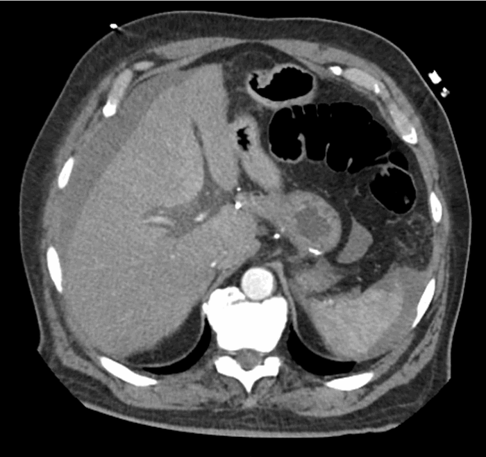
A 68-year-old male with no known history of cholecystitis or biliary disease presented to the emergency department with the complaint of diffuse epigastric pain. His medical history was significant for Roux-en-Y gastric bypass, noninsulin dependent diabetes, hypertension, acid reflux, and coronary artery disease status post a coronary artery bypass graft, for which he takes a daily baby aspirin. At that time, an evaluation for acute coronary syndrome was negative and he was discharged home with pain medication. There was no evaluation for abdominal pathology at this time. Three days later he again presented to the emergency department due to progression of his epigastric pain. At this time a diagnosis of acute cholecystitis was made by ultrasound with incidental finding of ascites (Figure 1). He had a significant leukocytosis (38.7 k/ul) and lactic acidosis (3.2 mg/dL). Given the severity of his illness, further imaging was obtained. A computed tomography (CT) scan demonstrated massively distended gallbladder with a blush of contrast in the lumen of the gallbladder concerning for active hemorrhage (Figure 2), as well as a significant amount of hemoperitoneum (Figure 3). The patient subsequently became hemodynamically unstable and was transported to a quaternary care center. He transiently responded to resuscitation with blood products but ultimately decompensated again before being emergently transported to the operating room. A midline exploratory laparotomy was performed that demonstrated a large amount of blood in the peritoneal cavity. All four quadrants were packed with lap sponges and old blood was evacuated. The bowel appeared to be decompressed. The gallbladder was found to be massively distended and filled with blood. One necrotic area had perforated, and blood was actively spraying into the abdomen. No other sources of bleeding were seen. The gallbladder was sharply opened and packed. A clear source of bleeding was not seen in the gallbladder but was controlled with packing. At this time pulses were lost and cardiac monitor with rhythm strips showed ventricular tachycardia. Despite attempts at resuscitation with advanced cardiac life support protocol, the patient ultimately expired, with a presumptive diagnosis of cardiogenic shock due to acute blood loss. An autopsy was not performed per patient family wishes.

This study was deemed exempt by University Hospitals Institutional Review Board.

Figure 1. Ultrasound image demonstrating distended gallbladder with thickened wall and ascites. Arrow head shows the distended gallbladder. Arrow shows ascites.
Figure 2. Distended gallbladder with intraluminal contrast suggesting active extravasation. Arrow shows a blush of contrast within gallbladder.
Figure 3. Large amount of hemoperitoneum throughout the abdomen.

Discussion

We performed literature review of hemorrhagic cholecystitis using a key word search via OVID medicine. Key words including “hemorrhagic cholecystitis”, “hemorrhage”, “acute cholecystitis” were used. Ten case reports and one retrospective review article were identified from 1987 to 2017 (Table 1). A total of 33 patients (including this case) were identified and their clinical characteristics described.

Author

Year Published

Article type

Number of patients

Outcome

Reference

Lai et al

2009

Case report

1

Discharged

3

Morris et al

2008

Case report

1

Discharged

5

Gremmels et al

2004

Case report

1

Discharged

6

Pandya et al

2008

Case report

1

Expired

4

Tavernaraki et al

2010

Case report

1

Expired

9

Hague et al

2010

Case report

3

Discharged

10

Stempei et al

1993

Case report

1

Discharged

1

Shope et al

2004

Case report

1

Discharged

7

Parekh et al

2010

Case report

2

Discharged

13

Shishida et al

2017

Case report

1

Discharged

2

Chin et al

1987

Retrospective review

19

Unknown

12

Table 1. Literature review

The average age was 63 and 70% were male. Risk factors identified include uremia, chronic kidney disease (CKD), atrial fibrillation, anticoagulation and/or antiplatelet therapy, chronic obstructive pulmonary disease (COPD) on chronic steroids, and blunt trauma. Among these patients, four patients had CKD and uremia1-3 and two were on anticoagulation therapy (Coumadin).4 Two patient were on aspirin5 and another one was on chronic steroid treatment for (COPD).6 Lastly, one patient had blunt abdominal trauma.7

Acute cholecystitis accounts for 3 to 10% of patients presented with abdominal pain.8 Hemorrhagic cholecystitis is a rare complication of this disease process. Despite the high incidence of acute cholecystitis, we were able to find only 32 hemorrhagic cases reported in the literature. Due to the rarity of the disease, hemorrhagic cholecystitis is not clearly defined in the literature and is used to describe either intraluminal or peritoneal hemorrhage secondary to acute cholecystitis. The degree of hemorrhage varies from asymptomatic to hemodynamically unstable. There exist no guidelines for diagnosis or treatment of this disease. Our experience with one such case, and our review of the literature, allowed us to attempt to generalize how best to diagnose and manage hemorrhagic cholecystitis.

These patients can present quite variably. In our collection of 33 patients, 100% of patients presented with abdominal pain. Some of them had nausea, vomiting and fever. All patients were hemodynamically stable at presentation. Signs of hemorrhage (hypotension, melena, lethargy, and anemia) developed in only 21% of the patients. Other laboratory abnormalities included leukocytosis in 56% of patients and hyperbilirubinemia was reported in 9% of patients. Patients rarely developed hemodynamically significant bleeding requiring close monitoring, adequate resuscitation, reversal of anticoagulation, and urgent intervention. Three cases resulted in death. Our case and the two other mortalities all had significant intra-abdominal bleeding and decompensated quickly. Signs of hemorrhage were present in less than a quarter of the patients in this series. The other patients in this series never developed hemodynamically instability, with 19 patients being diagnosed with hemorrhagic cholecystitis only by final pathology. Therefore, the hemodynamic stability should dictate the plan of management.

In the cases we reviewed, right upper quadrant ultrasound and CT scan of the abdomen and pelvis were the most common initial imaging studies in these patients. Ultrasound findings include distended gallbladder, wall thickening and pericholecystic fluid collection, with the most frequent diagnosis being acute cholecystitis. One study reported ultrasound finding of an intraluminal pulsatile mass.6 CT scans were performed in 11 patients, with only four of those demonstrating active intraluminal extravasation of contrast.4,5,9 A pseudoaneurysm of the cystic artery was present in three patients.10 Other findings suggestive of hemorrhage on CT scan included hemoperitoneum (one patient), blood clots in gallbladder (one patient), and heterogeneous intraluminal fluid (one patient) were described in three patients. Two patients had no significant CT findings. The initial imaging study of choice for biliary disease is ultrasonography.11 However, ultrasound does not appear to be ideal for differentiating between acute and hemorrhagic cholecystitis. The retrospective review study reported ultrasound findings on 19 acute cholecystitis patients with pathological findings of hemorrhage cholecystitis.12 They reported that ultrasound findings suggesting hemorrhagic cholecystitis included intraluminal membrane, focal gallbladder wall irregularities and nonshadowing, nonlayering intraluminal echoes.12 However, these findings were not present in the other 14 patients we reported here. Notably, because the retrospective study was published in 1987, CT scans might not have been widely available. We conclude that CT scan is useful in the diagnosis of hemorrhagic cholecystitis.

Most reports included the final pathology. Full thickness necrosis was found in 13 specimens. The remaining specimens showed features of cholecystitis and blood-filled gallbladders. All specimens demonstrated underlying cholecystitis.

We propose limiting the diagnosis of hemorrhagic cholecystitis to patients that present with signs and symptoms consistent with acute cholecystitis PLUS signs and symptoms of hemorrhage (hypotension, lethargy, melena, anemia), or imaging evidence of hemorrhage. Patients with hemodynamic stability can be managed successfully with laparoscopic cholecystectomy. Their outcomes were very good and all patients who underwent laparoscopic cholecystectomy were discharged home. However, exploratory laparotomy with open cholecystectomy was required in cases with hemodynamically instability. These outcomes were much poorer, as only 2 out of 5 patients who underwent laparotomy survived. While likely multifactorial, this poorer outcome can be attributed to a delayed diagnosis of this rare complication and these patients being at a more advanced stage in disease process at time of surgery. Interestingly, all five patients requiring laparotomy presented in stable condition. This suggests a window of time between presentation and clinical decompensation during which expedited evaluation and intervention could have been performed to allow a more favorable outcome. Hemodynamical instability in acute cholecystitis should be evaluate in the same manner as a trauma patient: assume bleeding until proven otherwise. Expedited resuscitation and urgent operative intervention are the only effective treatment; any delay will lead to poor outcome.

Conclusion

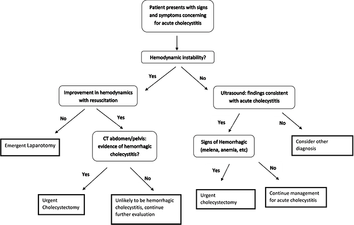
Hemorrhagic cholecystitis is an extremely rare but potentially deadly complication of acute cholecystitis. Prompt evaluation and intervention is required for patients with a concerning clinical presentation. Our proposed algorithm may help in the diagnosis and management of this disease entity (Figure 4).

Figure 4. A proposed algorithm for management of suspected hemorrhagic cholecystitis.

Lessons Learned

Hemorrhagic cholecystitis can range from a purely pathologic diagnosis to a fatal disease. A high index of suspicion is needed as this disease can be difficult to clinically diagnose. CT scan is a useful diagnostic tool, and prompt surgical intervention can be lifesaving in patients with hemorrhagic cholecystitis.

Authors

Bo Jiang, MD

Katherine Bingmer, MD
University Hospitals Cleveland Medical Center
Department of Surgery
Cleveland, OH

John Ammori, MD
University Hospitals Cleveland Medical Center
Department of Surgery
Cleveland, OH

Correspondence

John Ammori, MD
Associate Professor
Surgery, CWRU School of Medicine
11100 Euclid Ave
Department of Surgery
Cleveland, OH, 44106
Email: John.Ammori@uhhospitals.org
Phone: 216-844-1777

Disclosures

The authors have no conflicts of interest to report.

References

  1. Stempel LR and Vogelzang RL. Hemorrhagic Cholecystitis with Hemobilia: Treatment with Percutaneous Cholecystostomy and Transcatheter Urokinase. Journal of Vascular and Interventional Radiology. 1993;4:377-380.
  2. Shishida M, Ikeda M, Karakuchi N, Ono K, Tsukiyama N, Shimomura M, et al. Hemorrhagic Cholecystitis in a Patient on Maintenance Dialysis. Case Rep Gastroenterol. 2017;11:488-493.
  3. Lai YC and Tarng DC. Hemorrhagic acalculous cholecystitis: an unusual location of uremic bleeding. J Chin Med Assoc. 2009;72:484-7.
  4. Pandya R and O'Malley C. Hemorrhagic cholecystitis as a complication of anticoagulant therapy: role of CT in its diagnosis. Abdom Imaging. 2008;33:652-3.
  5. Morris DS, Porterfield JR and Sawyer MD. Hemorrhagic cholecystitis in an elderly patient taking aspirin and cilostazol. Case Rep Gastroenterol. 2008;2:203-7.
  6. Gremmels JM, Kruskal JB, Parangi S and Kane RA. Hemorrhagic cholecystitis simulating gallbladder carcinoma. J Ultrasound Med. 2004;23:993-5.
  7. Shope TR, Bass TL and Haluck RS. Laparoscopic management of traumatic hemorrhagic cholecystitis. JSLS. 2004;8:93-5.
  8. Kimura Y, Takada T, Kawarada Y, Nimura Y, Hirata K, Sekimoto M, et al. Definitions, pathophysiology, and epidemiology of acute cholangitis and cholecystitis: Tokyo Guidelines. J Hepatobiliary Pancreat Surg. 2007;14:15-26.
  9. Tavernaraki K, Sykara A, Tavernaraki E, Chondros D and Lolis ED. Massive intraperitoneal bleeding due to hemorrhagic cholecystitis and gallbladder rupture: CT findings. Abdom Imaging. 2011;36:565-8.
  10. Hague J, Brennand D, Raja J and Amin Z. Cystic artery pseudoaneurysms in hemorrhagic acute cholecystitis. Cardiovasc Intervent Radiol. 2010;33:1287-90.
  11. Shea JA, Berlin JA, Escarce JJ, Clarke JR, Kinosian BP, Cabana MD, et al. Revised estimates of diagnostic test sensitivity and specificity in suspected biliary tract disease. Arch Intern Med. 1994;154:2573-81.
  12. Chinn DH, Miller EI and Piper N. Hemorrhagic cholecystitis. Sonographic appearance and clinical presentation. J Ultrasound Med. 1987;6:313-7.
  13. Parekh J and Corvera CU. Hemorrhagic cholecystitis. Arch Surg. 2010;145:202-4.