Abstract
Background
A 41-year-old male with untreated human immunodeficiency virus (HIV) presented with tip appendicitis in the setting of newly diagnosed adult T-cell lymphoma.
Summary
Tip appendicitis is a variation of uncomplicated appendicitis with a variable clinical presentation and is sometimes diagnosed incidentally on imaging. The condition is usually found in the setting of an obstructing appendicolith. The overall prevalence of tip appendicitis is unknown to date. We present a case of tip appendicitis in an immunocompromised patient secondary to lymphadenopathy successfully managed medically.
Conclusion
Diagnosing and treating variations of acute uncomplicated appendicitis, such as tip appendicitis, is not well defined, particularly in immunocompromised patients. Our case report concludes that lymphoma can cause luminal obstruction leading to tip appendicitis and that select immunocompromised patients may be managed successfully nonoperatively.
Key Words
tip appendicitis; tip appendicitis with lymphoma; lymphoma
Case Description
Surgical management of abdominal pain due to acute appendicitis accounts for over 300,000 procedures annually in the United States.1 Historically, the diagnosis of acute appendicitis was based solely on clinical evaluation and physical exam. However, computed tomography (CT) and ultrasound (US) have become increasingly utilized, especially in clinically equivocal cases. While increased utilization of diagnostic imaging has helped to reduce the rate of negative appendectomies, it has also shed light on more subtle variations of appendiceal pathology for which established management guidelines do not exist. One such pathology is inflammation isolated to the distal appendix, usually with an obstructing appendicolith, often described as tip appendicitis.2
Tip appendicitis can present with nonspecific symptoms that can be mistaken for several variations of appendicitis, such as acute proximal appendicitis, unusual appendiceal location, perforations, or abscesses.2 Given such a wide differential diagnosis, imaging is more frequently used as an adjunct to clinical evaluation. This ultimately results in patients undergoing appendectomy based on clinical findings as well as positive imaging results. On diagnostic imaging, 39-57% of patients with tip appendicitis ultimately have a normal appendix on final pathology.3,6 Whether this radiological finding is, in fact, a clinical condition that would not have resolved without treatment is unknown. This uncertainty emphasizes the importance of the clinician's evaluation of the patient in deciding whether a patient needs surgery placed in context with abnormal imaging.
Appendicitis results from luminal obstruction, most frequently a fecalith, but can also be due to fecal stasis, lymphoid hyperplasia, neoplasms, undigested material, parasites, or previously ingested barium. This obstruction leads to rising pressures distally from gas production by bacteria and ongoing mucus secretion in the appendiceal lumen. As the appendix continues to distend, venous drainage decreases, resulting in mucosal ischemia and, ultimately, perforation. Stasis distal to the obstruction leads to bacterial overgrowth within the appendix, which can create a larger inoculum if perforation occurs.4
Here, we present a case of tip appendicitis in the setting of human T-cell leukemia/lymphoma virus-1 (HTLV-1) associated adult T-cell lymphoma (ATL). To our knowledge, this is the first reported case of a patient presenting with tip appendicitis in the setting of newly diagnosed ATL; we propose that a relationship exists between the two.
Our patient is a 41-year-old male with HIV untreated for one year who presented to the emergency room with a two-week history of worsening dysphagia. He also reported associated back pain, right lower quadrant (RLQ) and left lower quadrant (LLQ) abdominal pain, and 27-pound weight loss over the month. He has a history of dysphagia and was diagnosed with esophagitis and duodenal Strongyloides, treated successfully with Ivermectin.
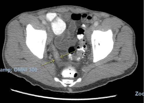
On physical examination, his vitals were within normal limits. He was very thin and had diffuse palpable lymphadenopathy in the cervical and inguinal regions and a large mobile left cheek mass. His abdominal exam was benign, with no anterior abdominal pain but rather bilateral flank pain. The patient's leukocytosis was 13,000 k/cm2, with the remainder of his labs within normal limits; the cluster of differentiation 4 (CD4) count was 2670, and the viral load was 1,391,823. HTLV-I/HTLV-II antibodies were positive. A CT of the abdomen and pelvis with oral and intravenous (IV) contrast was obtained, showing acute tip appendicitis (Figure 1) and extensive bulky gastrohepatic, periportal, peripancreatic, retroperitoneal/para-aortic, bilateral iliac chain, and bilateral inguinal lymphadenopathy (LAD) (Figure 2) with mild splenomegaly suggestive of lymphoma. A CT maxillofacial scan to evaluate his cheek mass also demonstrated extensive bulky cervical lymphadenopathy as well as an enlarged mandibular mass (Figure 3). Of note, the patient had a CT scan performed one year prior that showed an appendix with the same diameter but no local inflammation (Figure 4).
Figure 1. CT Scan: Tip Appendicitis in Pelvis. Published with Permission
Given the complexity of this patient, we took a multidisciplinary approach involving infectious disease, oncology, and radiology. The patient was admitted to the hospital, restarted on highly active antiretroviral therapy (HAART), and treated with IV antibiotics for the CT findings of tip appendicitis. Given his diffuse lymphadenopathy, he also underwent a cervical lymph node biopsy to provide a tissue diagnosis and further characterize his likely lymphoma.
Final pathology demonstrated atypical lymphoid proliferation suggestive of T-cell lymphoma, favoring Adult T-cell lymphoma/leukemia. Immunohistochemical stains revealed that the lymphoid cells are positive for all markers consistent with this diagnosis, further confirmed with flow cytometry.
The patient was successfully treated with IV antibiotics for the tip appendicitis. This was evidenced by the resolution of appendiceal tip hyperenhancement and filling with contrast on repeat CT scan one week after starting antibiotics (Figure 5 and Figure 6). He is being scheduled to initiate chemotherapy for the adult T-cell lymphoma.
Discussion
Acute appendicitis is among the most common surgically managed causes of abdominal pain.1 Up to 7% of the population will have appendicitis during their lifetimes, with a peak incidence between 10 and 30 years old.5 Presentation may include RLQ pain and tenderness, nausea, vomiting, anorexia in the setting of fever, and leukocytosis with a left shift.6 Given the risk of perforation and sepsis with delayed treatment, many surgeons have historically proceeded with appendectomy based on clinical findings and accepted a negative appendectomy rate (NAR) of 20% to 30%.6 Since the advent and widespread usage of imaging, CT scan and ultrasound have become the preferred diagnostic tools to diagnose and reduce the number of unnecessarily performed surgeries more accurately.
Inflammation localized to the distal portion of the appendix, the so-called tip appendicitis, is a rare disease process.2 While the true prevalence of tip appendicitis is unknown, a case study by Lim et al. reports the prevalence of clinically diagnosed and pathologically confirmed tip appendicitis to be as high as 5%.7 The clinical presentation associated with findings of tip appendicitis on imaging is variable. It may include RLQ pain and/or tenderness, anorexia, nausea and vomiting, fever, and leukocytosis with or without a left shift.6 Perforation of the appendiceal tip may occur. In contrast, data regarding perforation rates of tip appendicitis is limited. Lim et al. report perforation in 5/20 cases (25%), similar to the rate seen by Mazeh et al., who identified perforation in 4/26 cases (15%).6,7
Classic imaging findings suggestive of acute appendicitis include luminal dilation, wall thickening, and periappendiceal or RLQ inflammatory changes.2 Raja et al. found that from 1990 to 2007, as the proportion of adult patients having preoperative CT increased from 1% to 97.5%, the NAR decreased from 23% to 1.7%. This highlights the utility of advanced diagnostic imaging, especially in equivocal cases.8 While increased reliance on ever-improving diagnostic imaging has helped to reduce the NAR, it has also shed light on an increasing number of rare, more subtle variations of appendiceal pathology for which established management guidelines do not exist. One such pathology is described with classic findings isolated to the distal appendix, usually with an obstructing appendicolith, termed tip appendicitis.2
Despite the lack of formal diagnostic criteria for tip appendicitis, two studies provide a reasonable framework for diagnosis. A study by Leung et al. used the following criteria for diagnosis: a dilated distal appendix (≥6 mm) with the remainder of the appendix of normal caliber along with US findings of non-compressibility, hyperemia, wall-thickening, or loss of mural stratification; or CT findings of wall-thickening, mucosal and wall discontinuity, and mucosal hyperenhancement.3 A 2009 study on the diagnostic criteria for tip appendicitis necessitated several findings divided by location. In the proximal appendix, at least one of the following was required: normal caliber (<6 mm), luminal air, or luminal contrast. All the following were required for the distal appendix: dilation of at least 7 mm, wall thickening, and lack of luminal air or contrast.6 In addition, at least one of the following in the surrounding tissues: periappendiceal inflammatory changes or free fluid in the right lower quadrant.6
While imaging is valuable, imaging alone should not dictate clinical management. In a 2009 study of 18 patients with acute inflammation of the appendiceal tip, only seven (39%) underwent surgery and had confirmed acute appendicitis on final histopathology.6 Leung et al. observed similar findings, with 84% of patients with tip appendicitis on imaging (32 ultrasounds and 14 CT scans) ultimately not having appendicitis (positive predictive value = 16.4%) and a NAR of 57%.3 Therefore, finding tip appendicitis alone on imaging does not always correspond to positive appendiceal pathology following surgery. However, the study by Leung et al. did find that there were significant differences in regard to tachycardia, right lower quadrant tenderness, signs of peritonitis, and the presence or absence of polymorphonuclear neutrophilia when comparing positive to negative appendectomies.3 This stresses the importance of individualized clinical decision-making. If the surgeon feels the presentation is consistent with acute inflammation, they should proceed with operative intervention. However, if the findings are mild or the patient does not meet the criteria commonly used to identify classic acute appendicitis, observation and nonoperative management may be attempted.6 Nonoperative management of acute uncomplicated appendicitis is a controversial topic. While antibiotic treatment did not meet noninferiority criteria in the largest multicenter, open-label, randomized controlled trial conducted to date, 73% of patients with uncomplicated acute appendicitis were successfully treated (did not require appendectomy within one year) with broad-spectrum IV antibiotics followed by oral antibiotics.11
In this case, in addition to having uncontrolled HIV and acute tip appendicitis on imaging, the patient also had findings of diffuse LAD and mild splenomegaly, later confirmed to be HTLV-associated ATL. HTLV-1/2 infects 15-20 million individuals worldwide and is responsible for adult T-cell leukemia/lymphoma (ATLL).9 HTLV-1 and HIV-1 are common co-pathogens, and co-infection increases the risk of developing ATLL.10 ATLL can be classified into smoldering, chronic, lymphoma, and leukemic types.9 Patients who develop ATLL, especially the acute leukemic or lymphoma form, have a poor prognosis.9
To our knowledge, this is the first reported case of tip appendicitis in the setting of HTLV-associated ATL. We postulate that the lymphadenopathy caused by the HTLV-associated ATL is responsible for the patients' tip appendicitis, likely by obstruction of the distal appendiceal lumen, resulting in inflammation. The absence of appendicolith on imaging further supports our claim. Various neoplasms have been implicated in the development of acute appendicitis by similar processes, including mucinous cystadenoma, appendiceal carcinoid, primary appendiceal adenocarcinoma, and cecal carcinoma.2
Conclusion
Tip appendicitis is a variation of acute uncomplicated appendicitis. The diagnosis and treatment of appendicitis has evolved over the last few decades. Imaging as an adjunct to clinical evaluation has become standard practice, and antibiotics in unclear presentations have not shown inferior outcomes compared to surgical intervention. Our case demonstrates a unique cause of tip appendicitis in an immunocompromised patient successfully managed medically with IV antibiotics. Clinically, our patient did not have any classic findings of acute appendicitis, yet imaging revealed tip appendicitis. Through a multidisciplinary approach, given the diagnosis of ATL and lack of symptoms for appendicitis, we chose to manage the inflammation seen on imaging with IV antibiotics. In this case report, we conclude that tip appendicitis can be adequately and safely treated with IV antibiotics in immunocompromised patients. in select immunocompromised patients.
Lessons Learned
Tip appendicitis can be seen in patients with lymphadenopathy from lymphoma. Complex patients such as ours require a multidisciplinary approach but may still be safely treated with IV antibiotics.
Authors
Sharon C; Narula S; Sofia D; Lou I
Author Affiliation
The Brooklyn Hospital Center, Brooklyn, NY, 11201
Corresponding Author
Christina Sharon, MD
Department of Surgery
The Brooklyn Hospital Center
121 Dekalb Avenue, Ste. 8F
Maynard Building
Brooklyn, NY 11201
Email: tina.m.sharon@gmail.com
Disclosure Statement
The authors have no conflicts of interest to disclose.
Funding/Support
The authors have no relevant financial relationships or in-kind support to disclose.
Received: December 29, 2020
Revision received: May 11, 2021
Accepted: July 13, 2021
References
- Livingston EH, Fomby TB, Woodward WA, Haley RW. Epidemiological similarities between appendicitis and diverticulitis suggesting a common underlying pathogenesis. Arch Surg. 2011;146(3):308-314. doi:10.1001/archsurg.2011.2
- Gaetke-Udager K, Maturen KE, Hammer SG. Beyond acute appendicitis: imaging and pathologic spectrum of appendiceal pathology. Emerg Radiol. 2014;21(5):535-542. doi:10.1007/s10140-013-1188-7
- Leung B, Madhuripan N, Bittner K, et al. Clinical outcomes following identification of tip appendicitis on ultrasonography and CT scan. J Pediatr Surg. 2019;54(1):108-111. doi:10.1016/j.jpedsurg.2018.10.019
- Prystowsky JB, Pugh CM, Nagle AP. Current problems in surgery. Appendicitis. Curr Probl Surg. 2005;42(10):688-742. doi:10.1067/j.cpsurg.2005.07.005
- Addiss DG, Shaffer N, Fowler BS, Tauxe RV. The epidemiology of appendicitis and appendectomy in the United States. Am J Epidemiol. 1990;132(5):910-925. doi:10.1093/oxfordjournals.aje.a115734
- Mazeh H, Epelboym I, Reinherz J, Greenstein AJ, Divino CM. Tip appendicitis: clinical implications and management. Am J Surg. 2009;197(2):211-215. doi:10.1016/j.amjsurg.2008.04.016
- Lim HK, Lee WJ, Lee SJ, Namgung S, Lim JH. Focal appendicitis confined to the tip: diagnosis at US. Radiology. 1996;200(3):799-801. doi:10.1148/radiology.200.3.8756934
- Raja AS, Wright C, Sodickson AD, et al. Negative appendectomy rate in the era of CT: an 18-year perspective. Radiology. 2010;256(2):460-465. doi:10.1148/radiol.10091570
- Mahieux R, Gessain A. Adult T-cell leukemia/lymphoma and HTLV-1. Curr Hematol Malig Rep. 2007;2(4):257-264. doi:10.1007/s11899-007-0035-x
- Laher AE, Ebrahim O. HTLV-1, ATLL, severe hypercalcaemia and HIV-1 co-infection: an overview. Pan Afr Med J. 2018;30:61. Published 2018 May 28. doi:10.11604/pamj.2018.30.61.13238
- Salminen P, Paajanen H, Rautio T, et al. Antibiotic Therapy vs Appendectomy for Treatment of Uncomplicated Acute Appendicitis: The APPAC Randomized Clinical Trial. JAMA. 2015;313(23):2340-2348. doi:10.1001/jama.2015.6154


