Unsupported Browser
The American College of Surgeons website is not compatible with Internet Explorer 11, IE 11. For the best experience please update your browser.
Menu
Become a member and receive career-enhancing benefits

Our top priority is providing value to members. Your Member Services team is here to ensure you maximize your ACS member benefits, participate in College activities, and engage with your ACS colleagues. It's all here.

Become a Member
Become a member and receive career-enhancing benefits

Our top priority is providing value to members. Your Member Services team is here to ensure you maximize your ACS member benefits, participate in College activities, and engage with your ACS colleagues. It's all here.

Membership Benefits
ACS
Case Reviews in Surgery

Primary Acinar Cell Carcinoma of the Liver Complicated by Pancreatic Panniculitis

​A unique case of a female patient who presented with a primary acinar cell carcinoma of the liver complicated by pancreatic panniculitis.

July 8, 2021

Abstract

Background

Acinar cell carcinoma is a rare and aggressive neoplasm, almost exclusively of pancreatic origin. This carcinoma can rarely be complicated by subcutaneous panniculitis, termed “pancreatic panniculitis” that may serve as an early warning sign of the underlying disease. We present the unique case of a female patient who presented with a primary acinar cell carcinoma of the liver complicated by pancreatic panniculitis.

Summary

Our patient with BRCA 2 mutation and history of bilateral ovarian cancer at age 59 treated with bilateral salpingo-oophorectomy and seven cycles of carboplatin presented at age 67 with nausea, fatigue, anorexia, and tender, erythematous nodules in her lower extremities and left hand as well as polyarthritis. Comprehensive imaging and computed tomography (CT)-guided core biopsy revealed a single primary liver mass with features identical to acinar cell carcinoma of pancreatic type. Her nodules were initially presumed to represent erythema nodosum but later determined to be a presentation of pancreatic panniculitis given her underlying tumor and failure to resolve with a six-day course of topical steroids. The patient underwent an uncomplicated right hepatectomy to resect the mass, and her panniculitis resolved soon after the removal of the liver tumor. Final pathology revealed a stage II, pT2N0M0 acinar cell carcinoma. While pancreatic panniculitis secondary to pancreatic disease is well-documented, panniculitis resulting from primary liver disease is exceedingly rare. Moreover, the tumor, in this case, was of pancreatic cell differentiation, displaying the embryologic connection and similarities in oncologic differentiation between the pancreas and liver. The need for recognition of the diagnosis of pancreatic panniculitis as a foreshadowing of underlying disease, and impetus for further laboratory and imaging investigation, is underscored by this case report.

Conclusion

Pancreatic panniculitis is a rare complication of a rare cancer, acinar cell carcinoma. We present a unique case of primary acinar cell carcinoma of the liver complicated by pancreatic panniculitis, which resolved with surgical resection of the underlying tumor. It highlights the importance of recognizing non-resolving, tender, erythematous nodules as potentially representing pancreatic panniculitis. Furthermore, it underscores the need to consider both pancreatic and extrapancreatic disease in the differential diagnosis of pancreatic panniculitis.

Key Words

pancreatic panniculitis; acinar cell carcinoma; erythema nodosum; paraneoplastic


Case Presentation

Acinar cell carcinoma is most commonly a primary tumor of the pancreas, constituting 1 to 2 percent of pancreatic cancers.1 One of the known complications of acinar cell carcinoma is a subcutaneous panniculitis, thought to result from release of pancreatic enzymes such as lipase secondary to the disease, commonly termed “pancreatic panniculitis.” Such panniculitis symptoms are found in nearly 16 percent of patients with acinar cell carcinoma, which is the most common malignancy found in patients presenting with panniculitis.1 Extrapancreatic acinar cell carcinoma is a rare entity. It has been identified in locations including the liver, jejunum, colon, and stomach.25 We report the second case of pancreatic panniculitis associated with primary hepatic acinar cell carcinoma.

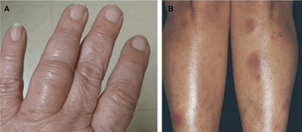
The patient is a 67-year-old Ashkenazi Jewish female with BRCA2 mutation and a history of bilateral ovarian cancer treated with bilateral salpingo-oophorectomy and seven cycles of carboplatin at age 59. Chronic conditions include diabetes, hypertension, hypothyroidism, and chronic obstructive pulmonary disease with 80-pack peryear smoking history. The patient presented with fatigue, nausea, anorexia, and tender, erythematous nodules on the lower extremities and left hand (Figure 1). Abdominal malignancy was high on the differential, especially given her BRCA2 mutation. She was found to have a large 11.1 cm necrotic right hepatic mass, noted on abdominal computed tomography (CT) scan, which was diagnosed at her initial institution as a neuroendocrine tumor. However, upon seeking a second opinion at our institution, pathology review of CT-guided core biopsy specimens later classified this as acinar cell carcinoma, and metastasis from a primary pancreatic tumor was initially presumed.

Figure 1. The patient’s left hand (A) and lower extremities (B) demonstrating pancreatic panniculitis, taken prior to surgery

The patient underwent endoscopic ultrasound (EUS), Gallium 68-DOTATATE positron emission tomography (PET, due to initial pathologic diagnosis of neuroendocrine tumor), whole-body nuclear medicine (NM) PET, magnetic resonance (MR) scan of the abdomen, chest CT, and mammogram that revealed no other lesions and no pancreatic tumor. The patient’s nodules were originally diagnosed as erythema nodosum by her dermatologist and treated with an ineffective six-day course of topical steroids. Lab values were notable for marked hyperlipasemia (>14,000 U/L, normal <50 U/L) upon presentation, white blood cell count of 13,900/µL (normal <10,000/µL) without eosinophilia, hemoglobin 9.9 g/dL (normal 12–16 g/dL). Liver function tests and tumor markers (AFP, CEA) were AST 28, ALT 18, ALP 168, total bilirubin 0.3, CEA <1, AFP <2, CA 19-9 <2. The remaining parameters were within normal limits. FDG PET scan of the whole body demonstrated the large liver lesion with no FDG-avid lymph nodes or metastases three days before the date of surgery (Figure 2).

Figure 2. Preoperative imaging: nuclear medicine positron emission topography (PET) scan demonstrating 9.6 cm necrotic lesion in the right lobe of the liver, segment 6/7; A) represents the scan without tracer; and B) demonstrates decreased fluoro-deoxyglucose (FDG) uptake in the area of the lesion compared to the surrounding liver, with a thick irregular rim of hypermetabolic activity—no extrahepatic lesions were noted

It was initially decided that the patient would be treated with resection alone. No staging laparoscopy was performed. After sterile preparation, an upper midline incision was made and extended rightward to create a hockey-stick incision. There was no evidence for local or peritoneal tumor spread on a thorough assessment of the abdomen. The liver tumor was identified with intraoperative ultrasound. Intraoperative ultrasound failed to detect a pancreatic mass. A right hepatectomy was performed, and the tumor was completely resected (Figure 3) with an estimated blood loss of 800 mL. The liver was not otherwise grossly abnormal. An incisional hernia from a prior operation was carefully reduced, and the hernia sac was sent to pathology, along with the gallbladder, which was removed.

Figure 3. Gross pathology: resected acinar cell carcinoma specimen from segment 6/7 of the liver

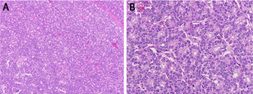
On gross pathological examination, the tumor was 15.2 cm in maximum diameter and confined to the liver. It was well-circumscribed but unencapsulated, with central cystic degeneration and areas of necrosis and hemorrhage. The surrounding liver parenchyma was grossly unremarkable. Histologic findings are shown in Figure 4. The tumor specimen stained positive for B-cell lymphoma/leukemia 10 (BCL10, an acinar cell marker) and trypsin, and negative for synaptophysin (neuroendocrine), chromogranin (neuroendocrine), and arginase-1 (a marker of liver differentiation). Immunohistochemistry findings can be seen in Figure 5. The final diagnosis was a moderately differentiated acinar cell carcinoma. The final pathologic stage was determined to be stage II, pT2N0M0. The adjacent parenchyma displayed no evidence of fibrosis.

Figure 4. Histology: liver mass on A) low and B) high magnifications. The liver mass was composed of tightly packed proliferations of cells forming acinar structures A) these structures resemble normal pancreatic acini in that they are formed by a single layer of cells with basally located nuclei; B) granular cytoplasm, arranging themselves around small lumens.

Figure 5. Immunohistochemistry: liver mass, showing A) positivity for B-cell leukemia/lymphoma 10 (BCL10); B) trypsin; and C) negative staining for synaptophysin

The patient received 1,618 mL of fluids and 2 units of packed red blood cells intraoperatively, and 1.5 L of normal saline on postoperative day 1 for rising creatinine. The patient developed symptoms of fluid overload, including 2+ pitting edema, shortness of breath, and 3-liter oxygen requirement, which normalized with furosemide diuresis. Within two days of surgery, the patient’s lipase level decreased from >14,000 U/L to 830 U/L, at which point her panniculitis was already resolving. One week post-operatively, her lipase had stabilized at 550 U/L, and her panniculitis was stable and improved from before resection.

Abdominal CT 65 days after surgery demonstrated no new evidence of malignancy. Gemcitabine and cisplatin were initiated at 96-day follow up as planned adjuvant treatment. The patient’s lipase had increased to 2,595 U/L by this point, raising concern for occult metastasis. Her panniculitis had completely resolved. Follow-up MRI 158 days after surgery confirmed two new liver lesions, measuring 2.6 cm and 1.3 cm (Figure 6). Cisplatin was replaced with oxaliplatin due to rising creatinine at this time. Gemcitabine and oxaliplatin were discontinued in favor of olaparib, an inhibitor of the enzyme poly ADP ribose polymerase (PARP), 223 days after surgery.

Figure 6. Postoperative imaging: magnetic resonance (MR) imaging of abdomen with contrast demonstrating two metastatic lesions in the left lobe of the liver located in segment two (A, 1.1 cm) and segment three (B, 2.7 cm)

At last clinical follow up, 209 days postoperatively, lipase levels had decreased to 475 U/L. There was no further evidence of panniculitis. There are no plans to treat with additional liver-directed therapy given evidence of metastasis, and the patient is managed by oncology for continued chemotherapy.

Discussion

Acinar cell carcinoma is an extremely aggressive cancer, with approximately half of patients harboring evidence of metastatic disease at the time of diagnosis.6 These neoplasms affect primarily adults with a mean age of 58 and show a striking predilection for men (male-to-female ratio 3.6:1).1 There have been seven reported cases of primary acinar cell carcinoma of the liver in the literature to date.7 Only one other reported case of primary hepatic acinar cell carcinoma associated with pancreatic panniculitis exists.7

Pancreatic panniculitis has largely been associated with pancreatic diseases, including pancreatitis, pancreatic carcinomas, or intraductal papillary mucinous neoplasm (IPMN).8 Nearly 45 percent of patients with pancreatic panniculitis show subcutaneous nodules before the underlying causal disease is recognized, indicating its utility as a valuable early sign of underlying malignancies and the need to conduct further laboratory and imaging studies.9 Although the pathophysiology remains uncertain, a biopsy of panniculitis will classically demonstrate “ghost cells,” or anucleate necrotic adipocytes with a basophilic granular material within their cytoplasm, as well as inflammation.10

In other reported cases of extrapancreatic acinar cell carcinoma, the disease is thought to originate from metaplastic, ectopic, or transdifferentiated pancreatic tissue, sharing biologic features with primary pancreatic acinar cell carcinoma.3 In fact, acinar cells have also been found to be able to transdifferentiate into hepatocytes.11 Furthermore, both hepatocellular carcinoma and acinar cell carcinoma are often driven by a dysfunctional Wnt/β-catenin pathway. Both demonstrate a surprisingly low frequency of KRAS mutations, and the most frequent chromosomal amplifications in both tumors are found on 1q, 8q, and 20q.12 Similar to previous reports of primary hepatic acinar cell carcinoma, the tumor, in this case, lacked biliary markers and hepatocellular differentiation, displayed positivity for trypsin and BCL10, demonstrated the presence of acini with apical cytoplasmic periodic acid-Schiff (PAS)-positivity, distinct acinar structures in non-desmoplastic stroma, absence of bile, and less high-grade nuclear features, distinguishing it from other primary liver tumors.

We describe the unique case of a patient with a primary liver tumor that showed features identical to that of pancreatic acinar cell carcinoma. Her case was complicated by pancreatic panniculitis, an extremely rare occurrence in the absence of primary pancreatic disease, which resolved with resection of her tumor. Our study adds to the previous literature in demonstrating that acinar cell carcinoma of the pancreatic type may also originate from the liver and can be readily distinguished from other primary liver neoplasms through histological and immunohistochemical features. It may cause a classic presentation of pancreatic panniculitis, an indicator of extremely high lipase levels, and possible presenting symptom of underlying malignancy, especially acinar cell carcinoma, of which practicing clinicians should be wary.

Conclusion

Pancreatic panniculitis is a rare complication of a rare cancer, namely acinar cell carcinoma. We present a unique case of extrapancreatic acinar cell carcinoma complicated by pancreatic panniculitis, which resolved with surgical resection of the underlying disease. It highlights the importance of considering extrapancreatic disease in the differential diagnosis of pancreatic panniculitis.

Lessons Learned

The presence of painful subcutaneous nodules that do not resolve with standard topical therapy should prompt consideration of the diagnosis of pancreatic panniculitis. This diagnosis is crucial, as it may further signify the presence of an extremely aggressive underlying malignancy, acinar cell carcinoma.

Authors

Jairath NKa; Jairath Ra; Westerhoff Mb; Nathan Ha

Author Affiliations

  1. University of Michigan, Department of Surgery, Ann Arbor, MI 48109
  2. University of Michigan, Department of Pathology, Ann Arbor, MI 48109

Corresponding Author

Neil Kunal Jairath
University of Michigan, Department of Surgery
Cancer Center, Floor B1, Reception E
1500 E Medical Center Drive SPC 5912
Ann Arbor, MI 48109
Phone: (734) 647-8902
E-mail: jairathn@med.umich.edu

Disclosure Statement

The authors have no conflicts of interest to disclose.

Acknowledgements

This manuscript was supported by unrestricted departmental support from the department of surgery and surgical oncology at the University of Michigan.

References

  1. Hruban R, Pitman M, Klimstra D. Tumors of the Pancreas. 4th Series, Fascicle 6. Washington; American Registry of Pathology, American Registry of Pathology and Armed Forces Institute of Pathology, Washington, DC; 2007. AFIP Atlas of Tumor Pathology.
  2. Agaimy A, Kaiser A, Becker K, et al. Pancreatic-type acinar cell carcinoma of the liver: a clinicopathologic study of four patients. Mod Pathol. 2011;24(12):1620-1626. doi:10.1038/modpathol.2011.127
  3. Lee H, Tang LH, Veras EF, Klimstra DS. The prevalence of pancreatic acinar differentiation in gastric adenocarcinoma: report of a case and immunohistochemical study of 111 additional cases. Am J Surg Pathol. 2012;36(3):402-408. doi:10.1097/PAS.0b013e318238369e
  4. Sun Y, Wasserman PG. Acinar cell carcinoma arising in the stomach: a case report with literature review. Hum Pathol. 2004;35(2):263-265. doi:10.1016/j.humpath.2003.09.020
  5. Makhlouf HR, Almeida JL, Sobin LH. Carcinoma in jejunal pancreatic heterotopia. Arch Pathol Lab Med. 1999;123(8):707-711. doi:10.5858/1999-123-0707-CIJPH
  6. Kitagami H, Kondo S, Hirano S, Kawakami H, Egawa S, Tanaka M. Acinar cell carcinoma of the pancreas: clinical analysis of 115 patients from Pancreatic Cancer Registry of Japan Pancreas Society. Pancreas. 2007;35(1):42-46. doi:10.1097/mpa.0b013e31804bfbd3
  7. Zundler S, Erber R, Agaimy A, et al. Pancreatic panniculitis in a patient with pancreatic-type acinar cell carcinoma of the liver--case report and review of literature. BMC Cancer. 2016;16:130. Published 2016 Feb 20. doi:10.1186/s12885-016-2184-6
  8. Marsh Rde W, Hagler KT, Carag HR, Flowers FP. Pancreatic panniculitis. Eur J Surg Oncol. 2005;31(10):1213-1215. doi:10.1016/j.ejso.2005.06.007
  9. Dahl PR, Su WP, Cullimore KC, Dicken CH. Pancreatic panniculitis. J Am Acad Dermatol. 1995;33(3):413-417. doi:10.1016/0190-9622(95)91385-8
  10. Laureano A, Mestre T, Ricardo L, Rodrigues AM, Cardoso J. Pancreatic panniculitis - a cutaneous manifestation of acute pancreatitis. J Dermatol Case Rep. 2014;8(1):35-37. Published 2014 Mar 31. doi:10.3315/jdcr.2014.1167
  11. Shen CN, Tosh D. Transdifferentiation of pancreatic cells to hepatocytes. Methods Mol Biol. 2010;640:273-280. doi:10.1007/978-1-60761-688-7_14
  12. Ghurburrun E, Borbath I, Lemaigre FP, Jacquemin P. Liver and pancreas: do similar embryonic development and tissue organization lead to similar mechanisms of tumorigenesis?. Gene Expr. 2018;18(3):149-155. doi:10.3727/105221618X15216414278706