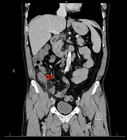
Furthermore, the specimen from the right hemicolectomy was with negative margins, while 6 out of 18 of the regional lymph nodes were positive for tumor invasion. Extranodal extension of the tumor was also noted as the right lateral abdominal biopsy was positive for scant tumor on the visceral peritoneum. Perineural invasion was also present. The prior incision site was negative for any malignancy.
Per the American Joint Committee on Cancer, the tumor presented as a clinical stage IIIC (cT4, cN2, cM0) and pathological stage IIIC (pT4a, pN2, cM0, G3). Due to positive nodal and extranodal involvement, the patient was a candidate for adjuvant chemotherapy. In addition to regular imaging and trending of tumor markers, he will also have a follow-up colonoscopy within one year to evaluate for any synchronous lesions or recurrence. Due to the aggressive and unpredictable nature of this tumor, he will require lifelong surveillance.
Discussion
GCCs are a rare biphasic group of neuroendocrine carcinomas that demonstrate mucinous and neuroendocrine properties.2 GCCs are known for their aggressive propensity, with most patients presenting with metastases at the time of diagnosis. They are also known to have a worse prognosis than the classic carcinoid counterparts and for their distinctive pattern of metastasis in that, they spread over the peritoneal surface and metastasize to lymph nodes.5,6
While previously believed to be an extension of colorectal carcinoma, advances in histologic analyses and genetic profiling have classified appendiceal carcinomas as a distinct entity from colorectal adenocarcinomas as well as from the classic carcinoid tumor of the appendix.7 However, confusion regarding the nomenclature of appendiceal goblet cell carcinoids still exists.4 For instance, while Tang et al.5 coined “adenocarcinoma ex-goblet cell carcinoid,” Taggart et al.8 coined “mixed goblet cell carcinoid-adenocarcinoma.” Nevertheless, Tang et al. classified GCCs into three groups based on histological findings: typical GCCs (group A); adenocarcinoma ex-goblet cell carcinoid, signet ring cell type (group B); and adenocarcinoma ex-goblet cell carcinoid, poorly differentiated carcinoma type (group C), which represents the worst prognosis of the groups.5 Consistent with the published literature, our patient with a Tang’s group C GCC tumor presented with nodal and extranodal involvement at the time of diagnosis, supporting the aggressive nature of GCCs.
Similar to its infrequent occurrence, preoperative diagnosis of GCCs is infrequent due to its mundane yet deceptive presenting symptoms, which are consistent with those of appendicitis. As a result, GCCs are typically identified post-appendectomy upon histological examination by the pathologist and require a second operation.9
Presently, primary appendiceal adenocarcinomas are managed with right hemicolectomy and lymphadenectomy, whereas the surgical management of typical carcinoid tumors is dictated by their size, grade, location, and invasion. Generally, in the absence of mesoappendiceal invasion, high grade, or location at the base of the appendix, carcinoids less than 1 cm in size can potentially be treated with an appendectomy, while carcinoids greater than 2 cm warrant right hemicolectomy.6
The proper surgical management of GCCs remains the subject of debate. However, the North American Neuroendocrine Tumor Society (NANETS)10 and the European Neuroendocrine Tumor Society (ENETS)11 guidelines currently recommend a right hemicolectomy as the appropriate surgical management for all GCCs, regardless of their histological type. Nevertheless, patients with increased perioperative morbidity who confer an unacceptable surgical risk, and patients who present with metastatic disease may not benefit from a right hemicolectomy and may be better suited for treatment with systemic chemotherapy.3,12 Tang et al. also recommended chemotherapy for both perforated GCCs and stage III and IV GCCs.5 Additionally, for women with GCCs, prophylactic bilateral oophorectomy is also frequently recommended.13
The National Comprehensive Cancer Network (NCCN) recommends appendiceal neoplasms be treated with systemic chemotherapy following their guidelines for colon cancer.14 Based on consensus opinion, chemotherapy regimens for GCCs also mirror that of colorectal adenocarcinoma. Chemotherapy regimens most commonly recommended include FOLFOX (5-FU, leucovorin, oxaliplatin) and FOLFIRI (5-FU, folic acid, irinotecan).3,12 Furthermore, a subset of patients with node-negative GCC or those with peritoneal carcinomatosis, may be candidates for cytoreductive surgery with hyperthermic intraperitoneal chemoperfusion (CRS-HIPEC).15,16
In summary, our case highlights the significance of examining the various presentations of the rare adenocarcinoma ex-goblet cell carcinoid tumor. Our patient’s initial presentation of a ruptured appendix was quite atypical due to the gradual onset of abdominal symptoms and his presenting age, which was consistent with the mean presenting age of GCC reported in the literature of 52 years.3 The information we reported in this case should alert physicians to the possibility of a diagnosis beyond appendicitis, even in the face of appendicitis-like symptoms in older patients with atypical presentations.
Conclusion
This case highlights a rare occurrence of poorly differentiated adenocarcinoma ex-goblet cell carcinoid following a common presentation of complicated appendicitis. Current recommendations suggest a right hemicolectomy with lymphadenectomy for all goblet cell carcinoids, regardless of the size of the primary tumor, depth of invasion, or histological grade. For tumors with nodal involvement, adjuvant chemotherapy is recommended.
Lessons Learned
Unlike carcinoid tumors of the appendix, which are considered a low-grade malignancy, goblet cell carcinoids are aggressive neoplasms that necessitate right hemicolectomy with lymphadenectomy followed by systemic chemotherapy stage III and IV disease.
Authors
Shaikh Sa; Patel M a ; Rosenthal AAb
Author Affiliations
- Nova Southeastern University, College of Osteopathic Medicine, Fort Lauderdale, FL 33314
- Memorial Regional Hospital, Division of Acute Care Surgery and Trauma, Hollywood, FL 33021
Corresponding Author
Andrew A. Rosenthal, MD, MBA, FACS
Division of Acute Care Surgery and Trauma
1150 N. 35th Avenue
Hollywood, FL 33021
Phone: (954) 265-5969
E-mail: anrosenthal@mhs.net
Meeting Presentation
South Florida Chapter, American College of Surgeons 2019 Annual Meeting
Best Case Report, Podium Presentation
Fort Lauderdale, FL
March 2019
Disclosure Statement
The authors have no conflicts of interest to disclose.
References
- Lietzén, Elina et al. “Appendiceal neoplasm risk associated with complicated acute appendicitis-a population based study.” International journal of colorectal disease vol. 34,1 (2019): 39-46. doi:10.1007/s00384-018-3156-x
- Reid, Michelle D et al. “Adenocarcinoma ex-goblet cell carcinoid (appendiceal-type crypt cell adenocarcinoma) is a morphologically distinct entity with highly aggressive behavior and frequent association with peritoneal/intra-abdominal dissemination: an analysis of 77 cases.” Modern pathology : an official journal of the United States and Canadian Academy of Pathology, Inc vol. 29,10 (2016): 1243-53. doi:10.1038/modpathol.2016.105
- Rossi, Roberta Elisa et al. “Goblet cell appendiceal tumors--management dilemmas and long-term outcomes.” Surgical oncology vol. 24,1 (2015): 47-53. doi:10.1016/j.suronc.2015.01.001
- Wang, Hanlin L, and Deepti Dhall. “Goblet or signet ring cells: that is the question.” Advances in anatomic pathology vol. 16,4 (2009): 247-54. doi:10.1097/PAP.0b013e3181a9d49a
- Tang, Laura H et al. “Pathologic classification and clinical behavior of the spectrum of goblet cell carcinoid tumors of the appendix.” The American journal of surgical pathology vol. 32,10 (2008): 1429-43. doi:10.1097/PAS.0b013e31817f1816
- Madani, Ariana et al. “Perforation in appendiceal well-differentiated carcinoid and goblet cell tumors: impact on prognosis? A systematic review.” Annals of surgical oncology vol. 22,3 (2015): 959-65. doi:10.1245/s10434-014-4023-9
- Jesinghaus, Moritz et al. “Appendiceal goblet cell carcinoids and adenocarcinomas ex-goblet cell carcinoid are genetically distinct from primary colorectal-type adenocarcinoma of the appendix.” Modern pathology : an official journal of the United States and Canadian Academy of Pathology, Inc vol. 31,5 (2018): 829-839. doi:10.1038/modpathol.2017.184
- Taggart, Melissa W et al. “Goblet cell carcinoid tumor, mixed goblet cell carcinoid-adenocarcinoma, and adenocarcinoma of the appendix: comparison of clinicopathologic features and prognosis.” Archives of pathology & laboratory medicine vol. 139,6 (2015): 782-90. doi:10.5858/arpa.2013-0047-OA
- Lee, K S et al. “Goblet cell carcinoid neoplasm of the appendix: clinical and CT features.” European journal of radiology vol. 82,1 (2013): 85-9. doi:10.1016/j.ejrad.2012.05.038
- Boudreaux, J Philip et al. “The NANETS consensus guideline for the diagnosis and management of neuroendocrine tumors: well-differentiated neuroendocrine tumors of the Jejunum, Ileum, Appendix, and Cecum.” Pancreas vol. 39,6 (2010): 753-66. doi:10.1097/MPA.0b013e3181ebb2a5
- Pape, Ulrich-Frank et al. “ENETS Consensus Guidelines for the management of patients with neuroendocrine neoplasms from the jejuno-ileum and the appendix including goblet cell carcinomas.” Neuroendocrinology vol. 95,2 (2012): 135-56. doi:10.1159/000335629
- Clift, Ashley K et al. “Goblet cell carcinomas of the appendix: rare but aggressive neoplasms with challenging management.” Endocrine connections vol. 7,2 (2018): 268-277. doi:10.1530/EC-17-0311
- Shenoy, Santosh. “Goblet cell carcinoids of the appendix: Tumor biology, mutations and management strategies.” World journal of gastrointestinal surgery vol. 8,10 (2016): 660-669. doi:10.4240/wjgs.v8.i10.660
- National Comprehensive Cancer Network, 2018, Colon Cancer, www.nccn.org/professionals/physician_gls/pdf/colon.pdf.
- McConnell, Yarrow J et al. “Cytoreductive surgery with hyperthermic intraperitoneal chemotherapy: an emerging treatment option for advanced goblet cell tumors of the appendix.” Annals of surgical oncology vol. 21,6 (2014): 1975-82. doi:10.1245/s10434-013-3469-5
- Radomski, Michal et al. “Curative Surgical Resection as a Component of Multimodality Therapy for Peritoneal Metastases from Goblet Cell Carcinoids.” Annals of surgical oncology vol. 23,13 (2016): 4338-4343. doi:10.1245/s10434-016-5412-z


