Unsupported Browser
The American College of Surgeons website is not compatible with Internet Explorer 11, IE 11. For the best experience please update your browser.
Menu
Become a member and receive career-enhancing benefits

Our top priority is providing value to members. Your Member Services team is here to ensure you maximize your ACS member benefits, participate in College activities, and engage with your ACS colleagues. It's all here.

Become a Member
Become a member and receive career-enhancing benefits

Our top priority is providing value to members. Your Member Services team is here to ensure you maximize your ACS member benefits, participate in College activities, and engage with your ACS colleagues. It's all here.

Membership Benefits
ACS
Case Reviews in Surgery

Traumatic Extrapleural Hematoma with Delayed Presentation Resulting in Tension Physiology Treated with Video-Assisted Thoracoscopy

Traumatic Extrapleural Hematoma with Delayed Presentation Resulting in Tension Physiology Treated with Video-Assisted Thoracoscopy

May 18, 2021

Abstract

Background

An adult male presented two days after falling onto his left side and sustaining multiple rib fractures, which resulted in a late-onset extrapleural hematoma with tension physiology.

Summary

Extrapleural hematomas (EPH) are a relatively rare complication of thoracic trauma with an incidence of 7.1 percent in one single-center study.1 The vast majority of these cases are associated with blunt mechanisms of injury.1 Rib fractures are present in up to 88 percent of cases, and the injury complex frequently includes pneumothoraces, hemothoraces, and pulmonary and chest wall contusions.1 Radiologic diagnosis of EPH is typically made within 24 to 48 hours of the initial injury, but less commonly a delayed presentation with interval development of an EPH occurs, leading to significant physiologic alterations and often resulting in surgical intervention.1,2 We present a unique case of a patient who suffered blunt chest trauma that was initially asymptomatic but then developed a late-onset EPH. This led to tension physiology requiring emergent video-assisted thoracoscopic drainage (VATS).

Conclusion

Delayed presentation of EPHs is a relatively rare complication of thoracic trauma in which an initially asymptomatic patient can present with life-threatening tension physiology several days after their injury. Minimally invasive techniques are effective for these patients, and unstable patients or those with tension physiology should be taken for VATS. Consideration should be given to initial extrapleural drainage before entering the thoracic cavity to more quickly relieve intrathoracic pressure and alleviate tension physiology if present.

Key Words

trauma; extrapleural hematoma; tension physiology; VATS


Case Description

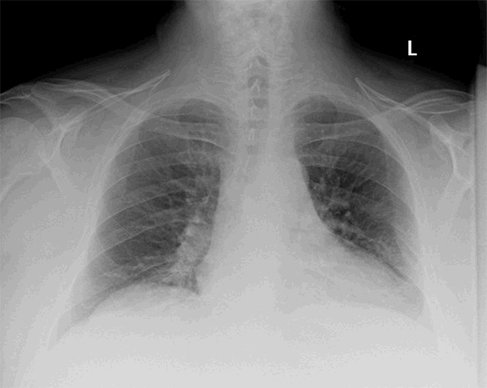
A 47-year-old man with a past medical history of hypertension on lisinopril and supraventricular tachycardia controlled with metoprolol and no history of blood thinning medications suffered a fall from standing and landed on his left side. He was initially managed at an outside hospital and discharged home with pain medications and a chest wall contusion diagnosis with no further evidence of worsening pulmonary function. A chest radiograph was performed as part of the workup that did not show any evidence of acute intrathoracic pathology (Figure 1).

Figure 1. Chest radiograph from the initial presentation at the outside hospital taken 48 hours before re-presentation.

Two days after the initial presentation, the patient developed worsening dyspnea and pain in the left chest. He re-presented to the outside hospital where a computed tomography (CT) scan identified closed, minimally displaced left-sided rib fractures four through seven and a large extrapleural hematoma without evidence of active extravasation (Figure 2). The trachea and mediastinum significantly deviated to the right. The patient was then transferred to our level I trauma center for further management.

Figure 2. A) CT scan of the chest (coronal view) from outside hospital at re-presentation noting a large extrapleural hematoma and a rightward shift of the mediastinal structures. B) CT scan of the chest (axial view)

Upon presentation, the patient’s temperature was 36.8°C, pulse 109 bpm, blood pressure 133/110 mmHg, and oxygen saturation 94 percent on room air. He was noted to be significantly dyspneic and diaphoretic with decreased breath sounds on the left on examination. The patient’s presentation, combined with a review of the CT scan, raised the concern for tension physiology. Therefore, the patient was taken emergently to the operating room for a left video-assisted thoracoscopy and evacuation of the hematoma. A tube thoracostomy was not placed in the trauma bay due to concern for further dissection of the parietal pleura from the chest wall and anticipated poor drainage of the already clotted hematoma. In the operating room, a dual-lumen endotracheal tube was placed upon induction. A 10 mm incision was made in the posterior axillary line at the fifth intercostals space, and the hematoma cavity was entered where extensive organized clot and some fresh blood was encountered and removed with suctioning. A second incision was then made at the same level in the anterior axillary line, and the pleural cavity was then accessed with the thoracoscope. This procedure allowed direct visualization of the remaining hematoma, which extended posteriorly. There was minimal serosanguinous fluid in the pleural space, confirming that the hematoma was extrapleural. The pleura was incised, and the remainder of the hematoma was drained through the pleural space. The hematoma cavity was examined, and no further signs of active bleeding were noted. There was no injury to the lung parenchyma. Two 32 French tube thoracostomies were placed in posterior basal and anterior apical positions. The patient remained intubated, was transferred to the surgical intensive care unit for further management and successfully extubated on postoperative day one. The anterior chest tube was removed on postoperative day two, and the posterior tube was removed on postoperative day three. He was then discharged home in good condition on postoperative day four. The patient was seen in the clinic for follow-up at one month with no intra-thoracic abnormalities noted on the chest radiograph (Figure 3) and full resolution of symptoms.

Figure 3. Chest radiograph at one month postdischarge during follow up clinic appointment

Discussion

Traumatic extrapleural hematomas were first described by Lipchik and Robinson in 19681 and further defined by Rashid et al. in 2000 in a case series of 34 patients.1 Here we present a case of a patient without any history of blood thinning medications who suffered blunt chest wall trauma, was initially asymptomatic, and then developed a late onset EPH leading to tension physiology necessitating emergent operative exploration and hematoma evacuation using a VATS approach. To our knowledge, this is the first report of tension physiology secondary to a delayed EPH.

Minimally invasive treatment options for EPHs include transcatheter arterial embolization (TAE) and VATS. TAE has been shown in multiple case reports to be a viable treatment modality for stable patients with EPH and has the benefit of avoiding the risks of a more extensive operation.5-7 In unstable patients and patients with an expanding EPH at risk for tension physiology, TAE is insufficient as it does not remove the hematoma nor relieve the pressure on the mediastinal structures.

The operative technique in prior case reports utilizing VATS for EPH involves entering the pleural cavity and then draining the hematoma intrapleurally with a partial pleurectomy if a connection was not already present.3,7,8 In contrast to this, the present case was managed with initial direct drainage of the extrapleural hematoma without entering the thoracic cavity, permitting immediate decompression of the hematoma, thereby stabilizing the patient. The pleural space could then be entered in a safe and controlled fashion. Further exploration then ensured there was no hemothorax component and allowed the remaining hematoma to be drained intrapleurally with a partial pleurectomy. In patients with tension physiology, this approach may decrease the time required to reduce the intrathoracic pressure and correct the underlying pathophysiology.

Conclusion

Delayed presentation of EPHs is a relatively rare complication of thoracic trauma in which an initially asymptomatic patient can present with life-threatening tension physiology several days after their injury. All patients, even asymptomatic, with rib fractures or at risk for missed rib fractures on initial imaging should have routine follow-up chest radiograph for monitoring their injuries as well as for rare complications such as EPHs. Minimally invasive techniques are effective for these patients, and unstable patients or those with tension physiology should be taken for VATS. Consideration should be given to initial extrapleural drainage before entering the thoracic cavity to more quickly relieve intrathoracic pressure and alleviate tension physiology if present.

Lessons Learned

Late-onset extrapleural hematomas may present with tension physiology following blunt chest trauma. Immediate operation is prudent and can be safely done with minimally invasive techniques.

Authors

Murray CR; Simon BJ; DeBusk MG

Author Affiliation

University of Massachusetts Medical School
Department of Surgery
Worcester, MA 01655

Corresponding Author

M. George DeBusk, MD, MSc, FACS
UMass Memorial Medical Center, S3-711
Department of Surgery
55 Lake Avenue North
Worcester, MA 01655
Phone: (508) 856-1168
E-mail: george.debusk@umassmemorial.org

Disclosure Statement

The authors have no conflicts of interest to disclose.

Acknowledgements

We would like to thank Karl Uy, MD, FACS, for providing technical and writing assistance.

References

  1. Lipchik, E O, and K E Robinson. “Acute traumatic rupture of the thoracic aorta.” The American journal of roentgenology, radium therapy, and nuclear medicine vol. 104,2 (1968): 408-12. doi:10.2214/ajr.104.2.408
  2. Rashid, M A et al. “Nomenclature, classification, and signficance of traumatic extrapleural hematoma.” The Journal of trauma vol. 49,2 (2000): 286-90. doi:10.1097/00005373-200008000-00016
  3. Masuda, Ryota et al. “Delayed hemothorax superimposed on extrapleural hematoma after blunt chest injury: a case report.” The Tokai journal of experimental and clinical medicine vol. 38,3 97-102. 20 Sep. 2013
  4. Pirzirenli, Mehmet Gökhan et al. “Extrapleural hematoma cases treated with video-assisted thoracoscopic surgery.” Ulusal travma ve acil cerrahi dergisi = Turkish journal of trauma & emergency surgery : TJTES vol. 21,5 (2015): 405-9. doi:10.5505/tjtes.2015.38768
  5. Morita, S et al. “Atypical extravasation of contrast medium in massive extrapleural haematoma due to cardiac massage without rib fracture.” Emergency medicine journal : EMJ vol. 26,10 (2009): 710. doi:10.1136/emj.2008.061739
  6. Gorospe, Luis et al. “Nonoperative Management of a Large Extrapleural Hematoma after Blunt Chest Trauma.” The Journal of emergency medicine vol. 51,2 (2016): 159-63. doi:10.1016/j.jemermed.2016.02.027
  7. Morita, Seiji et al. “Arterial embolization of an extrapleural hematoma from a dislocated fracture of the lumbar spine: a case report.” Scandinavian journal of trauma, resuscitation and emergency medicine vol. 17 27. 9 Jun. 2009, doi:10.1186/1757-7241-17-27
  8. Igai, Hitoshi et al. “Rapidly expanding extrapleural hematoma.” General thoracic and cardiovascular surgery vol. 56,10 (2008): 515-7. doi:10.1007/s11748-008-0284-1