Unsupported Browser
The American College of Surgeons website is not compatible with Internet Explorer 11, IE 11. For the best experience please update your browser.
Menu
Become a member and receive career-enhancing benefits

Our top priority is providing value to members. Your Member Services team is here to ensure you maximize your ACS member benefits, participate in College activities, and engage with your ACS colleagues. It's all here.

Become a Member
Become a member and receive career-enhancing benefits

Our top priority is providing value to members. Your Member Services team is here to ensure you maximize your ACS member benefits, participate in College activities, and engage with your ACS colleagues. It's all here.

Membership Benefits
ACS
Case Reviews in Surgery

Endoscopic Management of a Type III Paraesophageal Hernia Causing Gastric Outlet Obstruction in a Nonagenarian with Critical Aortic Stenosis

In the emergent setting, paraesophageal hernia (PEH) repairs are performed by open laparotomy with gastropexy and/or feeding tube placement, or laparoscopically, depending on the surgeon's expertise

December 4, 2020

Abstract

Background

In the emergent setting, paraesophageal hernia (PEH) repairs are performed by open laparotomy with gastropexy and/or feeding tube placement, or laparoscopically, depending on the surgeon's expertise. Recently, endoscopic techniques have been developed for high-risk patients. This case demonstrates the importance of preoperative risk assessment in determining which surgical intervention will provide the patient with the most symptom relief and the safest outcome.

Summary

A 90-year-old female with past medical history of symptomatic critical aortic stenosis, coronary artery disease, pacemaker, congestive heart failure, atrial fibrillation on Eliquis, pulmonary hypertension, hypothyroidism, GERD, and hiatal hernia presented to the emergency department with gastric outlet obstruction caused by a PEH. She was evaluated by cardiology, cardiothoracic surgery, internal medicine and anesthesia and was deemed prohibitive risk for definitive surgical repair. The patient successfully underwent endoscopic gastroduodenoscopy (EGD), endoscopic reduction of PEH, and gastropexy with insertion of a percutaneous endoscopic gastrostomy (PEG) tube. She had an uneventful postoperative course and was tolerating a mechanical soft diet prior to discharge. She was discharged to a skilled nursing facility on postoperative day four. At her two-week follow up, she was progressing very well without complication.

Conclusion

Endoscopic reduction and anterior gastropexy with PEG tube placement is a safe and effective method for management of incarcerated PEHs with gastric outlet obstruction in patients who are a prohibitive risk for surgery. This case helps in recognizing the importance of considering all surgical treatment options and weighing the risks and benefits of each when determining a plan of care for an elderly, high-risk patient.

Key Words

Paraesophageal hernia (PEH); gastric outlet obstruction (GOO), elderly; aortic stenosis; endoscopic; high-risk


Case Description

In the aging population, hiatal hernias are due to gradual enlargement of diaphragmatic hiatus. As the defect grows, the intra-abdominal esophagus, stomach, and or other organs can migrate above the diaphragm.1 Hiatal hernias are classified as type I–IV. Type I is a sliding hiatal hernia in which the gastroesophageal junction (GEJ) is displaced above the diaphragm.2 Type II–IV are paraesophageal hernias (PEHs). Type II results in migration of the stomach into the chest without GEJ, and in type III, the GEJ migrates with the stomach into the chest cavity..2 In a type IV PEH, the stomach is displaced into the chest along with other intraabdominal organs.2

Patients with a PEH may be asymptomatic for years, but typical presenting symptoms include chest pain, epigastric pain, heartburn, regurgitation, dysphagia, postprandial fullness, abdominal pain, vomiting, and anemia.13 PEHs have the potential to result in gastric volvulus, incarceration, strangulation, necrosis, and/or perforation; therefore, definitive surgical repair is recommended.1

Large type III and IV PEHs are most commonly found in the elderly population, more specifically females.3,4 Due to advanced age, patients have multiple medical comorbidities of varying severity. It is imperative that the operative risks are weighed against the risks of gastric incarceration or strangulation.1 Preoperative risk assessment is of great importance in this aging population, as some patients may deemed prohibitive risk for definitive surgical repair.1,4

The recommended surgical repair for PEH is laparoscopic reduction of the hernia, mobilization of GEJ, repair of diaphragmatic defect, and fixation of stomach within the abdomen with or without anti-reflux procedure such as a fundoplication.1,5,6 In the emergent setting, PEHs are typically repaired by open laparotomy with gastropexy and/or feeding tube placement, or laparoscopically, depending on the surgeon's expertise.3 That said, a few small studies have shown that laparoscopic or endoscopic reduction with gastropexy with or without laparoscopic assistance has been a successful means of hernia reduction and symptom relief in the high-risk, aging population.1,3,6,7 This case demonstrates the importance of preoperative risk assessment in determining which surgical intervention will provide the patient with the most symptom relief and the safest outcome.

In this current case, a 90-year-old female with past medical history of symptomatic critical aortic stenosis (AS), coronary artery disease, pacemaker, right and left heart catheterization, congestive heart failure, atrial fibrillation on Eliquis, pulmonary hypertension, GERD, and hiatal hernia presented to the emergency department with nausea, vomiting, epigastric pain, dysphagia to liquids with regurgitation, and weight loss for two weeks.

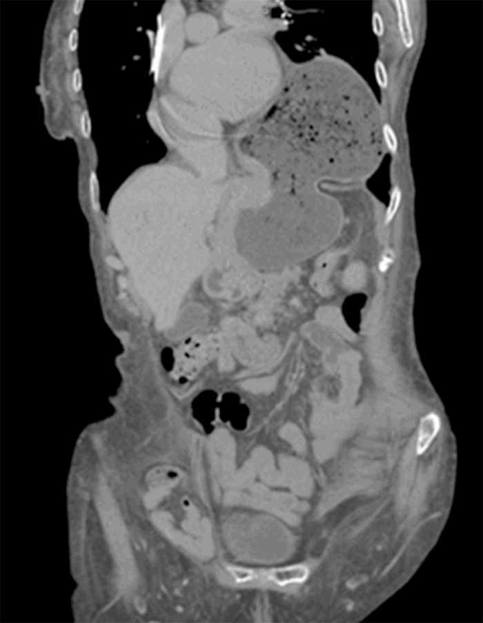
Computed tomography (CT) from outside facility performed earlier in the week revealed gastric outlet obstruction (GOO) caused by a massive type III PEH (Figure 1–4). She was subsequently discharged from this facility as she did not want surgical intervention and opted for hospice care. She presented to the ED later that week for persistent pain. At this time, it was decided to obtain a barium swallow instead of the repeat CT, demonstrating an intrathoracic stomach with concern for underlying gastric volvulus (Figure 5). Due to worsening obstructive symptoms, pain, and evidence of gastric outlet obstruction with possible gastric volvulus, it was recommended she undergo surgical intervention that day.

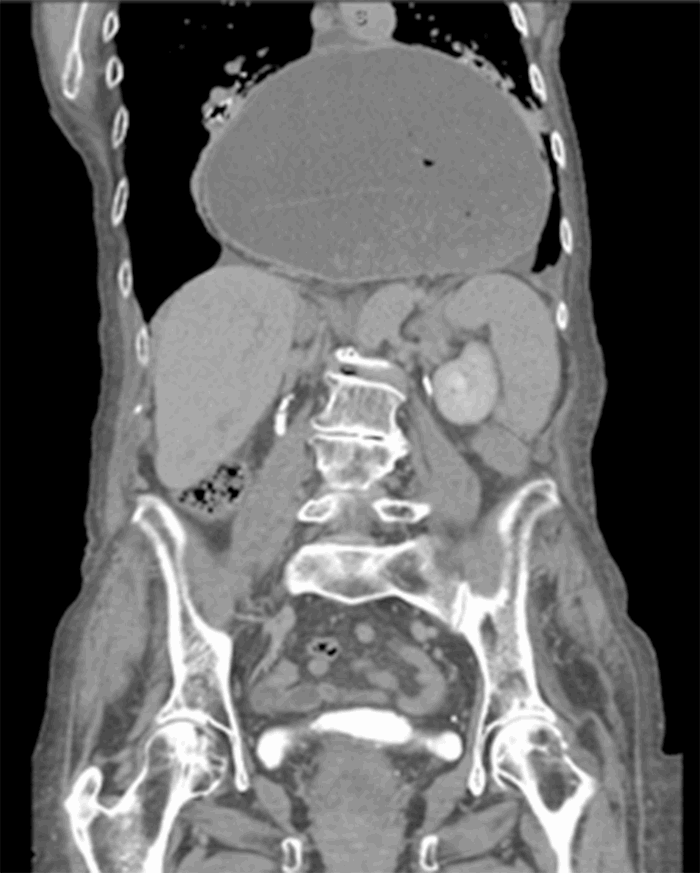
Figure 1. CT coronal view demonstrating large intrathoracic PEH.

Figure 2. CT coronal view demonstrating large intrathoracic PEH.

Figure 3. CT transverse mid-chest view.

Figure 4. CT transverse abdominal view.

Figure 5. Preoperative barium swallow demonstrating dilated intrathoracic stomach with GOO and concern gastric volvulus due to lack of gastric emptying.

Due to patient’s known history of severe AS, she was evaluated by cardiology, cardiothoracic surgery, internal medicine, and anesthesia, and she was deemed very high-risk for definitive laparoscopic surgical repair with an ASA grade assessment of IV and a procedure grade deemed emergent. Therefore, the decision was made to attempt an alternative, less invasive, and quicker approach via endoscopic reduction of the hernia with possible conversion to laparoscopy.

The patient was taken to the operating room and underwent endoscopic gastroduodenoscopy (EGD) and found to have large type III PEH with fluid and food that was removed via lavage. The PEH and volvulus was endoscopically reduced using the endoscope. The endoscope was retroflexed and, with gentle pressure, the board-based retroflexed scope was advanced, thereby reducing the hernia and detorsing the volvulus. The endoscope was then trans-illuminated through the stomach and abdominal wall in the epigastrium. Under endoscopic and direct visualization, a percutaneous endoscopic gastrostomy tube (PEG) was placed. Three T-fasteners were placed in triangulation around the PEG. A T-fastener consists of a metal bar located in the tip of an introducer needle with suture—this technique consists of passing the introducer needle percutaneously through the abdominal wall and into the stomach. The needle is then withdrawn, and the bar remains intraluminal. The external sutures are then fastened to the skin, securing the stomach up against the abdominal wall. The entire length of the procedure was 50 minutes. The patient remained stable throughout the entire procedure and was awakened from anesthesia and admitted to the ICU for ongoing care postoperatively. She developed asymptomatic bradycardia with pacemaker malfunction that was interrogated and adjusted by cardiology. She otherwise had an uneventful postoperative course, tolerated a mechanical soft diet, and was discharged to a skilled nursing facility on postoperative day four.

At her two-week follow-up, she was progressing very well without complication. A barium swallow was obtained (Figure 6), revealing a large, dilated, fluid-filled hiatal hernia with herniation of gastric fundus and proximal gastric body without evidence of obstruction and with prompt gastric emptying. No additional plans for repair of her hiatal hernia were made at this time. It was recommended that the patient follow up with cardiology to discuss TAVR prior to any elective repair.

Figure 6. Postoperative (three weeks) barium swallow demonstrating hiatal hernia with herniation of gastric fundus and proximal gastric body with prompt gastric emptying without evidence of obstruction.

Discussion

Patients in the tenth decade of life are at increased risk of developing large PEHs, incarceration, strangulation, and need for emergent intervention. In addition, they have a higher morbidity and mortality rate due to their multiple medical comorbidities.

The patient in this case had known symptomatic critical aortic stenosis, considered a high-risk index for cardiac complications during non-cardiac surgery (NCS).8 She had previously been seen by cardiology who recommended based on clinical guidelines by the American College of Cardiology/American Heart Association (ACC/AHA) that she undergo transcatheter aortic valve replacement (TAVR), which she refused.9

Patients with severe, symptomatic AS who underwent non-cardiac surgery were found to have high rates of cardiovascular complications, including myocardial infarct (MI), heart failure, ventricular arrhythmias, and intraoperative hypotension requiring pressor support, and death.9,10 It is recommended that they undergo valve replacement prior to surgery.9

On preoperative assessment by a multidisciplinary team, our patient was found to be at a very high risk for surgery. According to American Society of Anesthesiologist (ASA) she was deemed ASA class IV—a patient with severe systemic disease that is a constant threat to life. The Surgical Mortality Probability Model (S-MPM) estimates the 30-day mortality after NCS, and this model includes ASA score (I: 0 points, II: 2 points, III: 4 points, IV: 5 points, V: 6 points), procedure risk (low: 0 points, intermediate: 1 point, high: 2 points), and emergent (1 point) versus nonemergent intervention (0 points).11 Endoscopic procedures are considered low-risk and carry a less than 1 percent risk of cardiac death for NCS.11 The S-MPM class levels consist of class I (0–4 points with a mortality rate of less than 0.5 percent), class II (5–6 points with a mortality 1.5 to 4 percent), and class III (7–9 points with a mortality rate greater than 10 percent).11 The patient underwent a low-risk endoscopic approach with a S-MPM class II and a mortality rate of 1.5 to 4 percent. If she underwent an intermediate risk intraperitoneal surgery, she would be considered class III, and her associated mortality rate would increase to greater than 10 percent. Since our patient was classified as high-risk for intraoperative and postoperative complications, it was determined to attempt complete endoscopic reduction and gastropexy with laparoscopic assistance, if necessary.

It is important to note that endoscopic reduction of a PEH with gastropexy using PEG and T-fasteners does not repair the PEH (Figure 6). This technique does reduce the volvulus, relieve the GOO, and may reduce a portion of the herniated stomach, as seen in our case. The use of double PEG tube insertion for fixation of the stomach to the anterior abdominal wall has also been described in the literature as an effective treatment for reduction of symptomatic PEHs in this high-risk population.1 These techniques are ideal for elderly patients deemed high risk for surgery who need acute management of their PEH. These techniques are low-risk due to endoscopic approach and can be performed by surgeons in the emergent setting who do not necessarily have experience with formal laparoscopic or open PEH repairs. Additionally, these repairs do not require manipulation of the esophageal hiatus that renders elective definitive repair easier if needed once the patient is medically optimized and cleared for elective surgery.

Conclusion

A completely endoscopic reduction with gastropexy via PEG tube is a minimally invasive surgical option that should be seriously considered in patients that are deemed medically unfit to undergo a more extensive abdominal surgery. It is a safe and effective intervention that offers palliation of symptoms, decreased anesthetic and operative time, decreased mortality rate, decreased length of stay, and prevention of life-threatening complications.

Lessons Learned

This case recognizes the importance of considering all surgical treatment options when determining a plan of care for elderly, high-risk patients. It demonstrates the importance of considering preoperative risk assessment, risk factors. and life expectancy to provide the patient with the best surgical outcome and quality of life.

Authors

Johnson KR; Sellers WJ; Horsley RD

Corresponding Author

Kelly R. Johnson, DO
87 Mountain Rd.
Mountain Top, PA 18707
Phone: (845) 656-4818
E-mail: kelly.r.johnson9@gmail.com

Author Affiliation

Geisinger Community Medical Center
Department of General Surgery
1800 Mulberry St. Scranton, PA 18510

Meeting Presentation

2018 Annual Clinical Assembly-American College of Osteopathic Surgeons, Atlanta, Georgia, October 2018

Disclosure Statement

The authors have no conflicts of interest to disclose.

References

  1. Kercher KW, Matthews BD, Ponsky JL, et al. Minimally invasive management of paraesophageal herniation in the high-risk surgical patient. Am J Surg. 2001;182(5):510-514. doi:10.1016/s0002-9610(01)00760-7
  2. Straatman J, Groen LCB, van der Wielen N, et al. Treatment of paraesophageal hiatal hernia in octogenarians: a systematic review and retrospective cohort study. Dis Esophagus. 2018;31(7):10.1093/dote/doy010. doi:10.1093/dote/doy010
  3. Arevalo G, Wilkerson J, Saxe J. Acute Paraesophageal Hernia: Laparoscopic Repair With Adjunct T-Fastener Gastropexy for the High Operative Risk Patient. Surg Laparosc Endosc Percutan Tech. 2018;28(2):123-127. doi:10.1097/SLE.0000000000000509
  4. Higashi S, Nakajima K, Tanaka K, et al. Laparoscopic anterior gastropexy for type III/IV hiatal hernia in elderly patients. Surg Case Rep. 2017;3(1):45. doi:10.1186/s40792-017-0323-1
  5. Gangopadhyay N, Perrone JM, Soper NJ, et al. Outcomes of laparoscopic paraesophageal hernia repair in elderly and high-risk patients. Surgery. 2006;140(4):491-499. doi:10.1016/j.surg.2006.07.001
  6. Parker DM, Rambhajan AA, Horsley RD, Johanson K, Gabrielsen JD, Petrick AT. Laparoscopic paraesophageal hernia repair is safe in elderly patients. Surg Endosc. 2017;31(3):1186-1191. doi:10.1007/s00464-016-5089-1
  7. Yates RB, Hinojosa MW, Wright AS, Pellegrini CA, Oelschlager BK. Laparoscopic gastropexy relieves symptoms of obstructed gastric volvulus in highoperative risk patients. Am J Surg. 2015;209(5):875-880. doi:10.1016/j.amjsurg.2014.12.024
  8. Samarendra P, Mangione MP. Aortic stenosis and perioperative risk with noncardiac surgery. J Am Coll Cardiol. 2015;65(3):295-302. doi:10.1016/j.jacc.2014.10.051
  9. Kennon S, Archbold A. Expert Opinion: Guidelines for the Management of Patients with Aortic Stenosis Undergoing Non-cardiac Surgery: Out of Date and Overly Prescriptive. Interv Cardiol. 2017;12(2):133-136. doi:10.15420/icr.2017:20:2
  10. Calleja AM, Dommaraju S, Gaddam R, Cha S, Khandheria BK, Chaliki HP. Cardiac risk in patients aged >75 years with asymptomatic, severe aortic stenosis undergoing noncardiac surgery. Am J Cardiol. 2010;105(8):1159-1163. doi:10.1016/j.amjcard.2009.12.019
  11. Glance LG, Lustik SJ, Hannan EL, et al. The Surgical Mortality Probability Model: derivation and validation of a simple risk prediction rule for noncardiac surgery. Ann Surg. 2012;255(4):696-702. doi:10.1097/SLA.0b013e31824b45af
  12. Rendón-Medina MA, Ávalos-Abreu RO, Saucedo-Saldivar J, Sánchez-Tellez E, Garcia-Puig M. Laparoscopic treatment in Type IV Giant Paraesophagic Hernia and intestinal occlusion a case report. Int J Surg Case Rep. 2018;44:98-102. doi:10.1016/j.ijscr.2017.07.042