Unsupported Browser
The American College of Surgeons website is not compatible with Internet Explorer 11, IE 11. For the best experience please update your browser.
Menu
Become a member and receive career-enhancing benefits

Our top priority is providing value to members. Your Member Services team is here to ensure you maximize your ACS member benefits, participate in College activities, and engage with your ACS colleagues. It's all here.

Become a Member
Become a member and receive career-enhancing benefits

Our top priority is providing value to members. Your Member Services team is here to ensure you maximize your ACS member benefits, participate in College activities, and engage with your ACS colleagues. It's all here.

Membership Benefits
ACS
Case Reviews in Surgery

Gross Type C Tracheoesophageal Fistula with Duodenal Atresia, Distal Esophageal Stenosis, and Eosinophilic Esophagitis

Gross Type C Tracheoesophageal Fistula with Duodenal Atresia, Distal Esophageal Stenosis, and Eosinophilic Esophagitis

March 6, 2020

Abstract

Background

Esophageal atresia (EA) is a congenital malformation resulting in a blind-end esophagus within the mediastinum which most often presents with a tracheoesophageal fistula (TEF), an abnormal esophageal communication to the trachea. EA/TEF occurs in about 1 in every 2500-5000 births, and the most common variant, Gross Type C, accounts for about 85% of cases and is associated with a TEF from the trachea to the proximal end of the distal esophagus. Although EA/TEF does occur in isolation, many cases are associated with other birth defects such as the VACTERL (vertebral, anorectal, cardiac, tracheoesophageal, renal, limb) and CHARGE (Coloboma, Heart defects, Atresia of the choanae, Retardation, Genital hypoplasia, and Ear abnormalities) associations as well as genetic syndromes. Duodenal atresia (DA), congenital esophageal stenosis (CES), and eosinophilic esophagitis have been separately associated with tracheoesophageal fistula, but reports of all three conditions in the same patient are very rare.

Summary

In this case, a full-term baby was found to have Gross type C tracheoesophageal fistula and duodenal atresia. During surgical repair of the esophageal atresia, the lumen of the distal esophagus was found to be quite narrow. Distal esophageal stricture was confirmed by esophagram, and the narrowed segment was resected with primary anastomosis. She has developed eosinophilic esophagitis and required three esophageal dilations by the age of three years but is taking all food by mouth and gaining weight appropriately.

Conclusions

This case shows an atypical presentation of EA/TEF associated with both a distal CES and a duodenal atresia and underscores the importance of additional workup for unexpected intraoperative findings.

Key Words

Esophageal atresia, tracheoesophageal fistula, duodenal atresia, congenital esophageal stenosis


Case Description

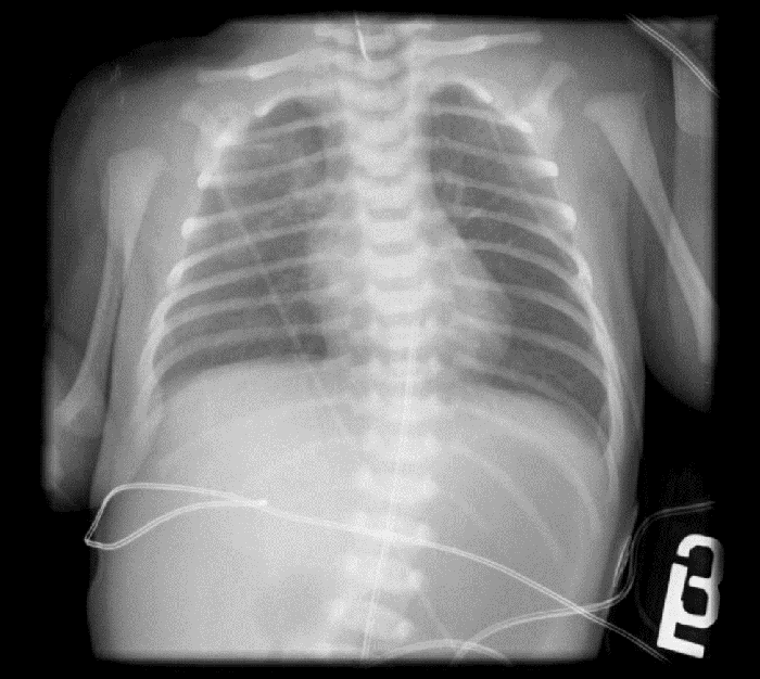
A 2725 g female infant with prenatal history of polyhydramnios was born at 39 weeks by Caesarean section for face presentation. She was placed on continuous positive airway pressure at delivery but quickly weaned to room air without evidence of respiratory distress. Initial x-ray demonstrated a coiled Replogle tube in the upper esophagus, an air-distended stomach, and a paucity of air in the distal gastrointestinal tract concerning for EA/TEF with DA (Figure 1).

Figure 1. Chest radiograph demonstrates a distended gas-filled stomach, and a Replogle tube in the esophagus which met resistance at the proximal esophageal pouch at the level of the clavicle.

Figure 1. Chest radiograph demonstrates a distended gas-filled stomach, and a Replogle tube in the esophagus which met resistance at the proximal esophageal pouch at the level of the clavicle.

Echocardiogram did not reveal abnormalities, and the baby was taken to the operating room urgently on day of life (DOL) 1 to prevent gastric rupture and aspiration. As our patient was initially stable on room air without massive gastric dilation, we elected to proceed first with fistula ligation and TEF repair with NGT decompression of the stomach; had her stomach been massively distended or if she had required more than a brief period of positive pressure ventilation, a gastrostomy tube would be required as an initial surgery to prevent gastric rupture, which has been reported in these circumstances. The baby became hypoxic following intubation, and bronchoscopy was performed which showed a large fistula one centimeter proximal to the carina. The endotracheal tube was maneuvered distal to the fistula. Unfortunately, the hypoxia persisted, and the decision was made to forego a thoracoscopic approach and proceed directly to a right posterolateral thoracotomy. The fistula was quickly controlled with a vessel loop, after which there was slow respiratory improvement. The fistula was ligated, the proximal pouch mobilized, and the posterior row of the esophageal anastomosis was performed. Repeated attempts were made to pass an 8 French feeding tube through the distal esophagus into the stomach, but each time resistance was met near the gastroesophageal junction. A 5 French feeding tube was successfully passed into the stomach. Duodenoduodenostomy might have been possible at this point; however, as we had achieved gastric decompression via NGT and the baby had earlier decompensated from a suspected aspiration, we elected to delay surgical correction of the DA. If we had failed to pass an NGT, we would have placed a gastrostomy tube with staged repair of the DA following recovery from the aspiration event and delayed repair of the CES in order to allow for esophageal healing, as it had been extensively mobilized during the TEF repair and we were concerned that a simultaneous resection of the second stricture would place the esophagus at risk for ischemia. The remainder of the case proceeded without event.

Our patient was extubated to room air the next day, and although she passed meconium after surgery, nasogastric tube (NGT) output remained high and on DOL 4 an upper gastrointestinal contrast study confirmed the suspected duodenal atresia (Figure 2). As the infant was now very stable, we returned to the operating room and she was intubated for surgery; intraoperatively she was found to have an annular pancreas and duodenal atresia. A duodenoduodenostomy was performed; bile was noted in the distal duodenum during repair. The remaining bowel was inspected, and no additional atresias were noted; she was extubated later the same day to nasal cannula which was weaned over the next 48 hours to room air.

Figure 2. Bedside contrast study postesophageal repair demonstrating failure of contrast to pass beyond into the proximal duodenum, consistent with duodenal atresia.

Figure 2. Bedside contrast study postesophageal repair demonstrating failure of contrast to pass beyond into the proximal duodenum, consistent with duodenal atresia.

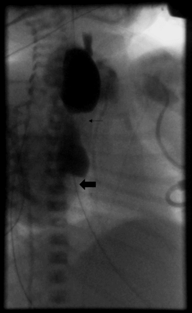
An esophagram was performed on DOL 8 to further evaluate the esophageal anastomosis and distal esophagus. The proximal esophagus was found to be dilated with passage of contrast through the esophageal anastomosis to a 2cm transition point in the distal esophagus with only slow passage of contrast into the stomach (Figure 3). There was no anastomotic leak. By DOL 10 she was tolerating full feeds by NGT and was discharged to an affiliate sub-acute pediatric care facility to allow for healing and weight gain prior to elective repair of the esophageal stricture.

Figure 3. Esophagram performed under fluoroscopy shows narrowing at the esophageal anastomosis (small arrow), with a second area of distal stricture (big arrow) with proximal distension. There was very slow passage of a small amount of contrast into the stomach.

Figure 3. Esophagram performed under fluoroscopy shows narrowing at the esophageal anastomosis (small arrow), with a second area of distal stricture (big arrow) with proximal distension. There was very slow passage of a small amount of contrast into the stomach.

Two months later, she electively underwent a resection of the narrowed distal esophagus with primary anastomosis via open right thoracotomy; after dissecting the esophagus above the diaphragm, we discovered a 1.5cm section of narrowed esophagus corresponding to the area seen on UGI. Additionally, a gastrostomy tube was placed in a laparoscopic fashion. Pathology of the resected short segment of esophagus showed a narrow lumen, mucosal reactive changes, and mild chronic inflammation.

Feeding therapy was started and by 4 months of age, she was taking all feeds by mouth. She has undergone three esophageal dilations at age 1.5 years, 2.5 years, and 3 years of age, with endoscopic biopsies each time consistent with eosinophilic esophagitis. She has gained weight appropriately.

Discussion

Roughly half of EA/TEF cases present with other major malformations, most commonly cardiac, skeletal, and anal abnormalities.1-8 Some 5 to 10% of cases have underlying chromosomal abnormalities, most commonly trisomy 18 and trisomy 21. In a study of 463 infants presenting with EA/TEF over an 18-year span, 107 (23%) had two or more defects described in the VACTERL association.8 Of the 107 patients in that cohort, 17 had chromosomal syndromes. Non-VACTERL abnormalities were common in the remaining 90 patients in that study, including an 8.9% incidence of duodenal atresia. In another study of 99 infants presenting with EA/TEF, there were 2 cases of duodenal atresia.7 This is a much higher frequency than the general population, where duodenal atresia has a reported incidence of 1 in 10,000 births.9 The exact pathological association between duodenal atresia and EA/TEF remains unclear, but both conditions are substantially more common in patients with trisomy 21.

Congenital esophageal stenosis has been associated in infants presenting with EA/TEF, separate from narrowing at the esophageal anastomosis. In fact, CES does appear to be diagnosed and repaired earlier in infants with distal CES and EA/TEF compared to CES alone.6 In a retrospective review of 187 children with EA/TEF who underwent postoperative esophagram,10 22 patients (12%) were diagnosed with distal CES, defined by "a narrowing below the anastomosis and above the GE junction with proximal dilation and no evidence of acquired cause of stenosis." The age at diagnosis ranged from 1 week to 10 years; half of the children had clinically significant symptoms such as dysphagia, postprandial emesis, and food/foreign body impaction. The etiology of the stenosis was classified by histology as either a tracheobronchial remnant (most common) or fibromuscular hypertrophy; as in other series, esophageal balloon dilation was either ineffective or resulted in perforation and the authors recommended surgical resection as first line therapy. In addition to EA/TEF and CES, one patient also had duodenal atresia and imperforate anus, one of only two cases of EA/TEF with CES and DA that could be found in our search of the literature.10,11 The esophageal stenosis in our case was present at the time of the first operation on DOL1; accordingly, we felt comfortable with the diagnosis of CES rather than an acquired cause such as reflux, although perhaps in utero reflux due to the duodenal atresia could have contributed.

EE has a reported prevalence of 1/10,000; yet reports of EE associated with EA are not infrequent; once case series reported an incidence of 17%.12 EE was diagnosed months to years following repair and was associated with more reflux symptoms and a higher incidence of fundoplication. Gastroesophageal reflux disease is also common after EA repair, especially in "wide gap" cases requiring more extensive mobilization of the esophagus; in patients refractory to medical therapy, endoscopy with biopsy should be sought to exclude EE.

Conclusions

This case shows an atypical presentation of EA/TEF associated with a distal CES, eosinophilic esophagitis, and a duodenal atresia, and underscores the importance of additional workup for unexpected intraoperative findings.

Lessons Learned

Although esophageal atresia with trachea-esophageal atresia and duodenal atresia were diagnosed from the initial plain film, it was necessary in this case to conduct the surgical repair in stages. Gastric decompression is a priority to prevent rupture of the stomach. Congenital esophageal stenosis is associated with esophageal atresia; if the infant is gaining weight on tube feeds, repair can be done electively at a higher weight.

Authors

Chris T Laird, MD
University of Maryland School of Medicine
Department of General Surgery
Baltimore, MD

Natalie A O'Neill, MD
University of Maryland School of Medicine
Department of General Surgery
Baltimore, MD

Kimberly M Lumpkins, MD, FACS
University of Maryland School of Medicine
Department of Pediatric Surgery
Baltimore, MD

Eric D Strauch, MD, FACS
University of Maryland School of Medicine
Department of Pediatric Surgery
Baltimore, MD

Corresponding Author

Chris Laird, MD
22 S Greene Street
Department of General Surgery, 8th floor
Baltimore, MD 21201
Phone: (443) 904-0070
Email: claird@som.umaryland.edu

Disclosure Statement

The authors have no financial disclosures. No funding or grant support. This report does not contain any personal information that could lead to the identification of the patient.

References

  1. Depaepe A, Dolk H, Lechat MF. The epidemiology of tracheo-oesophageal fistula and oesophageal atresia in Europe. EUROCAT Working Group. Arch Dis Child. 1993Jan;68(6):743–8. PMCID: PMC1029365
  2. Neilson IR, Croitoru DP, Guttman FM, Youssef S, Laberge J-M. Distal congenital esophageal stenosis associated with esophageal atresia. J Pediatr Surg. 1991;26(4):478–82. https://doi.org/10.1016/0022-3468(91)90999-A
  3. Newman B, Bender TM. Esophageal atresia/tracheoesophageal fistula and associated congenital esophageal stenosis. Pediatr Radiol. 1997Nov;27(6):530–4. PMID: 9174027 DOI: 10.1007/s002470050174
  4. Kim S-H, Kim H-Y, Jung S-E, Lee S-C, Park K-W. Clinical Study of Congenital Esophageal Stenosis: Comparison according to Association of Esophageal Atresia and Tracheoesophageal Fistula. J Pediatr Gastroenterol Nutr. 2017;20(2):79.
  5. Geneviève D, Pontual LD, Amiel J, Sarnacki S, Lyonnet S. An overview of isolated and syndromic oesophageal atresia. Clin Genet. 2007Feb;71(5):392–9. doi: 10.1111/j.1399-0004.2007.00798.x
  6. Holcomb GW, Ashcraft KW. Ashcrafts pediatric surgery. London: Saunders Elsevier; 2014.
  7. Stoll C, Alembik Y, Dott B, Roth M-P. Associated malformations in patients with esophageal atresia. Eur J Med Genet. 2009;52(5):287–90. doi:10.1016/j.ejmg.2009.04.004
  8. Carli D, Garagnani L, Lando M, Fairplay T, Bernasconi S, Landi A, et al. VACTERL (Vertebral Defects, Anal Atresia, Tracheoesophageal Fistula with Esophageal Atresia, Cardiac Defects, Renal and Limb Anomalies) Association: Disease Spectrum in 25 Patients Ascertained for Their Upper Limb Involvement. J Pediatr. 2014;164(3).
  9. Choudhry MS, Rahman N, Boyd P, Lakhoo K. Duodenal atresia: associated anomalies, prenatal diagnosis and outcome. Pediatr Surg Int. 2009;25(8):727–30.
  10. Yoo HJ, Kim WS, Cheon J-E, Yoo S-Y, Park K-W, Jung S-E, et al. Congenital esophageal stenosis associated with esophageal atresia/tracheoesophageal fistula: clinical and radiologic features. Pediatr Radiol. 2010Oct;40(8):1353–9.
  11. McCann, F., Michaud, L., Aspirot, A., Levesque, D., Gottrand, F. and Faure, C. (2014). Congenital esophageal stenosis associated with esophageal atresia. Dis Esophagus. 28(3), pp.211-215.
  12. Krishnan U. Eosinophilic Esophagitis in Children with Esophageal Atresia. European J Pediatr Surg. 2015; 25: 336–344.