Figure 5. Foreign body pathology specimen.
Discussion
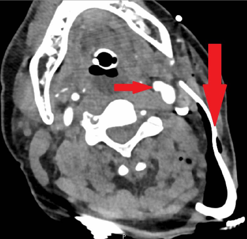
Penetrating injury to the neck is a serious and potentially life threatening injury. These injuries put patients at risk for decompensation during the first key steps of the Advanced Trauma Life Support approach to trauma: Airway, Breathing, Circulation, Disability. In today’s clinical practice there is still the widespread utilization of the classical “zone” approach to characterizing the injury to the neck. This approach divided the neck into three regions and was used to help determine the next steps in management. Zone one and three injuries led to the use of endoscopy and angiography with zone two injuries requiring mandatory surgical neck exploration. In our case, there was a clear injury to zone three, the use of the step-wise zone approach to neck trauma was utilized and complimented by CT angiography once the patient was assessed to be hemodynamically stable and followed closely by surgery for foreign body removal rather than the initial described approach of initial angiography.1
Zone three injuries are demarcated by the angle of the mandible to the skull base containing trachea, esophagus, jugular veins, carotid arteries, vertebral arteries, cranial nerves IX-XII and spinal cord. A patient presenting with a penetrating neck injury is assessed in the trauma bay as being hemodynamically stable or unstable. Furthermore, these stable patients are classified as being symptomatic or asymptomatic. All unstable patients no matter to what zone the injury is localized are taken to the operating room for surgical neck exploration. The hemodynamically stable patients with an isolated injury whether symptomatic or not can be managed with initial evaluation by CT of the neck and chest which drives selective non-operative management.
Historically, a protocol for mandatory neck exploration of any penetrating neck trauma was advocated, given the cost of missing a critical injury is high. However, this was shown to be inefficient since more than half of explorations resulted in no major injury found.2 Guidelines now suggest selective nonoperative management as an appropriate treatment pathway.3
Currently for surgical assessment there is growing support for the “No Zone” evaluation of neck injuries as contrasted with the traditional zone method described above. Typically, as with many protocols, expert surgeons utilize them as guidelines and implement anecdotal experience with intuition when establishing an intervention course.4 With the advent of CT angiography, hemodynamically stable patients can be evaluated with a radiographic study in combination with the standard trauma physical examination, regardless of the zone of injury thus changing the zone approach towards guiding management.
The current “no zone” algorithm for penetrating neck trauma focuses on identifying hard signs of injury and assessing patient stability rather than looking to the zone of injury for management. It has been shown that using CT angiography in a stable patient with a penetrating neck injury has led to better outcomes and is favorable in comparison to the zone approach.5 In addition, research has demonstrated that traditional “zoning” of the neck injury is not entirely accurate since almost 20 percent of zone II injuries were shown to enter zone I or III internally;6 however the more concerning issues are those zone I or III injuries that enter zone 2 which automatically would be considered surgical. The zone approach is still useful for helping clinicians quickly and effectively describe the anatomic location of the injury.7
Conclusion
The traditional classification of neck injuries based on zone gives a rapid and efficient algorithm for intervention. However, with high-resolution cross-sectional imaging, the trend is moving away from treatment algorithms based on neck zones. The utilization of imaging in conjunction with the focused trauma physical exam can provide an effective roadmap for the trauma surgeon and help ensure the best outcome for the patient by creating a clearer indication for when invasive intervention is required. Numerous published case series and reviews have supported the utility of the “no zone approach” to penetrating neck trauma.9,10,11
Lessons Learned
Penetrating neck trauma is an evolving field and while the traditional neck zones can be used as a framework for anatomic consideration, a shift toward a more patient-focused “no zone” technique is appropriate in the setting of more advanced diagnostic imaging.
Authors
Ryan B. Fransman, MD
Department of Surgery, The Johns Hopkins Hospital, Baltimore, MD
Faris K. Azar, MD
Department of Surgery, The Johns Hopkins Hospital, Baltimore, MD
William Mallon, MD
Department of Surgery, The Johns Hopkins Hospital, Baltimore, MD
Abhinav Nalluri, BS
Johns Hopkins University School of Medicine, Baltimore, MD
Joseph V. Sakran, MD, MPH, MPA
Department of Surgery, The Johns Hopkins Hospital, Baltimore, MD
Elliott R. Haut, MD, PhD
Department of Surgery, The Johns Hopkins Hospital, Baltimore, MD
Correspondence Author
Elliott R. Haut, MD, PhD, FACS
Department of Surgery
The Johns Hopkins University School of Medicine
1800 Orleans St, Zayed 6107c
Baltimore, MD
410-502-3122
ehaut1@jhmi.edu
Disclosure
The authors whose names are listed immediately above certify that they have NO affiliations with or involvement in any organization or entity with any financial interest (such as honoraria; educational grants; participation in speakers' bureaus; membership, employment, consultancies, stock ownership, or other equity interest; and expert testimony or patent-licensing arrangements), or non-financial interest (such as personal or professional relationships, affiliations, knowledge or beliefs) in the subject matter or materials discussed in this manuscript.
References
- Gracias VH. Computed tomography in the evaluation of penetrating neck trauma: a preliminary study. Arch Surg. 2001. Nov;136(11):1231–5.
- Muller R. Results of mandatory exploration for penetrating neck trauma. World J. Surg. 1994 Nov-Dec; 18(6):917–9.
- Tisherman SA, Bokhari F, Collier B, Cumming J, Ebert J, Holevar M, Kurek S, Leon S, Rhee P. Clinical practice guideline: penetrating zone II neck trauma. J Trauma. 2008 May;64(5):1392–405.
- Grosh JD. Management of penetrating neck injuries. The controversy surrounding zone II injuries. Surg Clin North Am. 1991 Apr; 71(2): 267–96
- Shiroff AA. Penetrating neck trauma: a review of management strategies and discussion of the 'No Zone' approach. The American surgeon. 2013-01;79:23.
- Bell RB, Osborn T, Dierks EJ, Potter BE, Long WB. Management of Penetrating Neck Injuries: A New Paradigm for Civilian Trauma. J Oral Maxillofac Surg. 2007 Apr;65(4):691–705.
- Long WB. Management of Penetrating Neck Injuries: a new paradigm for civilian trauma. J Oral Maxillofac Surg. 2007 Apr; 65 (4): 691–705.
- Roepke C, Benjamin E, Jhun P, Herbert M. Penetrating Neck Injury: What’s In and What’s Out? Ann Emerg Med 2016; 67: 578–580.
- Low GM, Inaba K, Chouliaras K et al. The use of the anatomic ‘zones’ of the neck in the assessment of penetrating neck injury. Am Surg 2014; 80: 970–974.
- Samorn P, Kritayakirana K, Sriussadaporn S, Pak-art R et al. Selective Management of Penetrating neck injuries using “no zone” approach. Injury 2015 Sep; 46(9) 1720–5.
- Ooi E, Stew B, Nowicki JL. Penetrating neck injuries: a guide to evaluation and management. Ann R Coll Surg Engl. 2017 Oct 19: 1–6.


