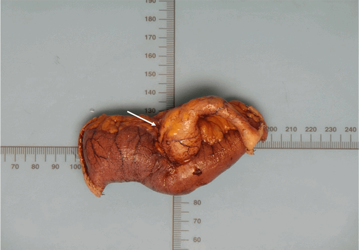
Pathology revealed a well-differentiated, 1.5 cm T4b (based on small bowel invasion) colonic type appendiceal adenocarcinoma (CAA) of the appendix with negative tumor margins. The patient underwent colonoscopy to evaluate for evidence of coexisting neoplasms that showed no evidence of colorectal neoplastic processes. After discussion of his treatment options, the patient elected to undergo right hemicolectomy for appropriate nodal staging. Pathology on the hemicolectomy specimen showed no evidence of metastatic disease or lymph node involvement, and the patient’s recovery was uneventful. He has a T4bN0M0 CAA and is following up with medical oncology.
Discussion
SBOs are most commonly due to adhesions and abdominal hernias; SBO secondary to mechanical appendiceal obstruction are an extremely rare phenomenon.7,8 Cases in the literature that exist usually involve an acute appendiceal inflammatory process and evidence of small bowel strangulation—neither was present in the patient of this current report. Of the existing 23 cases in the literature, 16 involved a twisted appendiceal tourniquet that encircled involved small bowel.7 An additional seven cases described obstruction secondary to a mucocele, only one of which was due to a malignant process.7 To our knowledge, there are no reports of CAA presenting with an SBO in the literature as of the time of this writing.
Our patient had two CT scans over his hospital courses that showed evidence of obstruction with a transition point in the RLQ. Intraoperatively, the distal tip of the appendix was observed to be adherent to, and apparently infiltrating, the distal ileum. Given the intermittent and self-resolving nature of the patient’s SBOs, it is likely that the described case represented a partial obstruction with the adherent appendix functionally behaving like an adhesion.
Evidence-based management for CAA is limited, and treatment guidelines are generally based on colon adenocarcinoma.3 Tumors smaller than 2 cm not involving the appendiceal base and without evidence of mesoappendix invasion or lymph node involvement are sometimes managed with appendectomy alone.4 For tumors larger than 2 cm, lymph node involvement, or mesoappendix invasion, right hemicolectomy is generally favored and is what was performed in the case of our patient.4 In patients with lymph node involvement, adjuvant therapy with 5-flurourical/leucovorin is warranted.4 Additionally, all patients should undergo complete colonoscopy to identify any coexisting colorectal neoplasms.
Due to their rarity, risk factors for APMNs have not yet been identified.4 The most common manifestation of APMN is acute appendicitis, and diagnosis is often made through the pathology report on the appendectomy specimen.4 Of note, colonoscopy is rarely able to detect nonmetastatic APMNs.9 As such, it is of critical importance to be aware of the imaging characteristics of the most common APMNs (please see below). The three most common types of APMN are CAA, mucinous type adenocarcinoma (MAA), and appendiceal neuroendocrine tumors (ANETs).
MAAs are the most common APMN, representing roughly 37 percent of all APMNs.1,4 MAAs characteristically show up on CT scans as appendiceal mucoceles with extra-appendiceal mucin being highly suggestive of a malignant process.10 These tumors are differentiated from CAA by their production of large amounts of mucin. Both MAAs and CAAs are subtypes of epithelial adenocarcinomas. Patients with advanced MAA often present with abdominal distention secondary to pseudomyxoma peritonei (PMP) caused by a ruptured primary tumor leaking mucinous fluid into the peritoneum. Interestingly, despite its rarity, MAA is actually by far the most common cause of PMP, despite PMP being classically associated with ovarian neoplasms.3,4
The second most common APMNs are CAAs, representing 25 percent of APMNs. CAAs appear on CT imaging as a soft tissue mass with possible regional adenopathy.4 CAAs are generally believed to arise by the same adenoma-carcinoma sequence as colon cancer, and, as mentioned, management of these neoplasms mirrors colorectal cancer guidelines. There is considerably less literature describing CAA than MAA, possibly due to CAA’s lack of unique characteristics or presentation.
ANETs can be either benign or malignant, and they represent 20 percent of all APMNs.1 ANETs are extremely difficult to diagnose radiographically, and, due to their small size, they are often missed. If visible, ANETs may be seen as a small mass on the distal appendix with regional adenopathy.4 ANETs commonly present in younger patients around the fourth decade of life, and occur more commonly in women than men.1,4 Neuroendocrine tumors are classically associated with carcinoid syndrome; however, this is an uncommon manifestation of ANETs, present only approximately 5 percent of the time and usually only after liver metastasis.4
Conclusion
The appendix is a rare but documented cause of mechanical bowel obstruction. In this article, the authors reported the case of a 70-year-old man presenting with one month of recurrent SBOs ultimately found to have a CAA.
Lesson Learned
APMNs are often insidious and detected incidentally. In patients with abdominal pain and evidence of SBO, APMN should be on the differential after more likely etiologies have been ruled out.
Authors
Sinensky A; Patel K; McKeown D; Marks JA
Corresponding Author
Joshua A. Marks, MD, FACS
Division of Acute Care Surgery
Department of Surgery
Thomas Jefferson University
1100 Walnut Street, Suite 702
Philadelphia, PA 19107
Phone: (215) 955-2165
E-mail: joshua.marks@jefferson.edu
Author Affiliation
Thomas Jefferson University
Department of Surgery
Division of Acute Care Surgery
Philadelphia, PA 19107
Disclosure Statement
The authors have no conflicts of interest to disclose.
References
- McCusker ME, Coté TR, Clegg LX, Sobin LH. Primary malignant neoplasms of the appendix: a population-based study from the surveillance, epidemiology and end-results program, 1973-1998. Cancer. 2002;94(12):3307-3312. doi:10.1002/cncr.10589
- Marmor S, Portschy PR, Tuttle TM, Virnig BA. The rise in appendiceal cancer incidence: 2000-2009. J Gastrointest Surg. 2015;19(4):743-750. doi:10.1007/s11605-014-2726-7
- Kelly KJ. Management of Appendix Cancer. Clin Colon Rectal Surg. 2015;28(4):247-255. doi:10.1055/s-0035-1564433
- Leonards LM, Pahwa A, Patel MK, Petersen J, Nguyen MJ, Jude CM. Neoplasms of the Appendix: Pictorial Review with Clinical and Pathologic Correlation. Radiographics. 2017;37(4):1059-1083. doi:10.1148/rg.2017160150
- Gündoğar Ö, Kımıloğlu E, Komut N, et al. Evaluation of appendiceal mucinous neoplasms with a new classification system and literature review. Turk J Gastroenterol. 2018;29(5):533-542. doi:10.5152/tjg.2018.17605
- Connor SJ, Hanna GB, Frizelle FA. Appendiceal tumors: retrospective clinicopathologic analysis of appendiceal tumors from 7,970 appendectomies. Dis Colon Rectum. 1998;41(1):75-80. doi:10.1007/BF02236899
- Komo T, Kohashi T, Hihara J, et al. Intestinal obstruction caused by low-grade appendiceal mucinous neoplasm: A case report and review of the literature. Int J Surg Case Rep. 2018;51:37-40. doi:10.1016/j.ijscr.2018.08.001
- Malý O, Páral J. Appendicitis as a rare cause of mechanical small-bowel obstruction: A literature review of case reports. Int J Surg Case Rep. 2016;29:180-184. doi:10.1016/j.ijscr.2016.10.065
- Trivedi AN, Levine EA, Mishra G. Adenocarcinoma of the appendix is rarely detected by colonoscopy. J Gastrointest Surg. 2009;13(4):668-675. doi:10.1007/s11605-008-0774-6
- Tirumani SH, Fraser-Hill M, Auer R, et al. Mucinous neoplasms of the appendix: a current comprehensive clinicopathologic and imaging review. Cancer Imaging. 2013;13(1):14-25. Published 2013 Feb 22. doi:10.1102/1470-7330.2013.0003