Unsupported Browser
The American College of Surgeons website is not compatible with Internet Explorer 11, IE 11. For the best experience please update your browser.
Menu
Become a member and receive career-enhancing benefits

Our top priority is providing value to members. Your Member Services team is here to ensure you maximize your ACS member benefits, participate in College activities, and engage with your ACS colleagues. It's all here.

Become a Member
Become a member and receive career-enhancing benefits

Our top priority is providing value to members. Your Member Services team is here to ensure you maximize your ACS member benefits, participate in College activities, and engage with your ACS colleagues. It's all here.

Membership Benefits
ACS
Case Reviews in Surgery

Colonic Paralytic Ileus in COVID-19 Patients

We present three patients with COVID-19 infection who developed colonic paralytic ileus.

December 18, 2020

Abstract

Background

We present three patients with COVID-19 infection who developed colonic paralytic ileus.

Summary

Our three patients presented with respiratory and generalized symptoms suggestive of coronavirus disease 2019 (COVID-19) infection. This diagnosis was subsequently confirmed by polymerase chain reaction testing. All patients developed acute respiratory distress syndrome (ARDS) necessitating intubation and mechanical ventilation. They were all uniformly coagulopathic, with elevated serum fibrinogen and D-dimers. During their hospitalization, the patients were found to have colonic ileus, with one progressing to bowel ischemia and requiring operative intervention. The other two patients were successfully managed nonoperatively.

Conclusion

As data continue to accumulate on the systemic manifestations of the COVID-19 virus, clinicians should be aware of the development of what appears to be a unique colonic paralytic ileus that could progress to bowel ischemia.

Key Words

COVID-19; colonic paralytic ileus; bowel ischemia


Case Description

With more than 20 million confirmed coronavirus disease 2019 (COVID-19) infections worldwide, including 5.1 million in the United States alone, the COVID-19 pandemic has become one of the most devastating pandemics of the modern age in terms of spread, morbidity, and mortality.1,2 In the United States and its territories, the Centers for Disease Control and Prevention (CDC) reported more than 163,000 deaths as of August 13, 2020.2

While the disease affects the respiratory system primarily, with the rapid development of acute respiratory distress syndrome (ARDS) in severe cases, front-line clinicians are quickly realizing that COVID-19 is a systemic disease with renal, hematological, and gastrointestinal manifestations. In this series from our institution, we report that among the critically ill COVID-19 patients, some develop a severe paralytic colonic ileus resembling colonic pseudo-obstruction (Ogilvie syndrome), with the risk of progression into full-thickness necrosis necessitating emergent surgical intervention.

Case 1

A 69-year-old male with a history of chronic obstructive pulmonary disease, active tobacco use, and coronary artery disease presented with a one-day history of cough, hemoptysis, and epistaxis. There was no fever, shortness of breath, chest pain, nausea, vomiting, abdominal pain, diarrhea, or myalgias. The patient denied recent travel or exposure to known sick contacts. On examination, he was afebrile, with an oxygen saturation of 97 percent on room air. A friable oral cavity/oropharyngeal mass was also noted.

On laboratory examination, his white blood cell count (WBC) was 6900/uL, with lymphopenia of 19.3 percent (normal: 22 to 44 percent). D-dimer was initially normal at 458 ng/mL (normal: <500 ng/mL) but continued to rise, with a peak at 3750 ng/mL. Serum fibrinogen level was elevated to 1034 mg/dL (normal: 150–400 mg/dL). Tests for respiratory syncytial virus and influenza were negative. Chest X ray showed bilateral linear lung atelectasis and scarring. CT angiogram of the chest showed signs of emphysema and chronic bronchitis (Figure 1a). A nasopharyngeal swab was sent for polymerase chain reaction (PCR) testing and was positive for COVID-19.

Figure 1. Case 1 CT imaging (a) CT angiogram of the chest showed signs of emphysema and chronic bronchitis; (b) CXR consistent with acute respiratory distress syndrome; (c) abdominal X-ray showing generalized large bowel dilatation

The patient was admitted for resuscitation and monitoring. He was started on hydroxychloroquine and azithromycin. His hospital course was complicated by coronary vasospasm that led to a new onset chest pain, ST segment elevation on electrocardiogram, bradycardia, and hypotension, followed by progressive hypoxemic respiratory failure and ARDS requiring admission to the ICU and intubation on the seventh hospital day (Figure 1b) as well as shock requiring vasopressors. He improved clinically in the ICU, and his ST elevation resolved, so he did not undergo percutaneous coronary intervention or received antiplatelet therapy. He was, however, started on heparin for deep venous thrombosis prophylaxis. Despite an initial ileus, he started having bowel movements and tolerated full nasogastric nutrition. Two weeks after admission, he started having a new and significant abdominal distension. An abdominal X ray was obtained showing severe gaseous distension of the colon measuring 12 cm at the cecum level, with gas noted within the rectum and no obstructive pattern (Figure 1c). The WBC was normal at 11,400 /uL. A rectal tube was placed, and the patient immediately passed a large amount of flatus and 600–1000 mL liquid brown stool; his abdominal distention improved. The subsequent clinical course of this patient has been published previously in the Annals of Surgery.3 Briefly, twenty-four hours after his last abdominal X ray, he developed a new leukocytosis to 22,000/uL, his abdominal distention worsened again, and a repeat abdominal X ray showed worsening colonic gaseous distension up to 13.5 cm at the cecum, now with signs of bowel wall pneumatosis. The patient was taken urgently to the operating room for an exploratory laparotomy. Intraoperatively, there was diffuse dilatation of the entire colon with patchy necrosis of the cecum, necessitating a total abdominal colectomy and an end ileostomy. His colon pathology showed ischemic large bowel with focal transmural necrosis.3 He was transferred to the ICU in critical condition. He subsequently developed acute kidney injury and was started on continuous veno-venous hemodialysis. Due to multiple end-organ dysfunction and poor prognosis, the family elected to transition him to comfort measures only as consistent with his previously expressed wishes. He subsequently died.

Case 2

A 49-year-old male presented with a one-week history of malaise, fever, cough, and shortness of breath. On examination, he was febrile to 103⁰F, hypoxemic to 82 percent, tachypneic, and hypotensive. Chest X ray showed bilateral patchy opacities. He quickly developed ARDS requiring intubation and mechanical ventilation. His nasopharyngeal swab for PCR testing for COVID-19 was positive. His WBC was notable for lymphopenia of 11.0 percent (normal: 22–44 percent). D-dimers and fibrinogen were elevated at 995 ng/mL (normal: <500 ng/mL) and 811 mg/dL (150–400 mg/dL), respectively. His ARDS continued to deteriorate, so he was placed on veno-venous extracorporeal membrane oxygenation (ECMO) after failing prone positioning and inhaled nitric oxide treatment. The patient was on heparin infusion while on ECMO.

The patient was noted not to be having bowel movements, and his abdomen became progressively distended. An abdominal X ray showed generalized colonic distension up to 8 cm (Figure 2a). Repeat X rays documented progression of the colonic distention up to 10 cm (Figure 2b). His WBC count was 4000 /uL and serum lactate was 0.9 mg/dL. A presumptive diagnosis of acute colonic pseudo-obstruction versus COVID-19 related paralytic colonic ileus was made. A rectal tube was placed, resulting in the evacuation of a large amount of flatus. Soap and water enema were also administered along with an aggressive bowel regimen. Neostigmine use was considered but never administered. Repeat KUB a few days later showed progressive improvement and resolution of the colonic distension (Figure 2c). The patient subsequently continued to improve, was decannulated from ECMO, extubated, and was transferred to the floor. After he was weaned off ECMO, he was started on subcutaneous Lovenox. The patient was subsequently discharged to a post-acute care facility.

Figure 2. Case 2 CT imaging (a) abdominal X-ray showing generalized colonic distension; (b) abdominal X-ray showing progressive colonic distension; (c) abdominal X-ray showing resolution of colonic distension

Case 3

A 43-year-old female with a history of hypertension, hyperlipidemia, and reactive airway disease presented with a one-week history of cough, fever, shortness of breath, and myalgia. She had a history of exposure to a close contact with fever and cough. On examination, she was febrile with a temperature of 102⁰F, tachycardic, had an oxygen saturation <94 percent on 4 L/min of oxygen, and had bilateral lung crackles. On laboratory evaluation, the WBC count was 8,400/uL with lymphopenia at 7.8percent (normal: 22 to 44 percent), D-dimers were elevated at 1361 ng/mL as well as fibrinogen at 618 mg/dL, C-reactive protein at 145.5 mg/L [normal: <8 mg/dL], and erythrocyte sedimentation rate (59 mm/h [normal: 0–22 mm/h]). A nasopharyngeal swab for PCR testing was positive for COVID-19.

The patient was admitted to the telemetry floor and started on hydroxychloroquine, atorvastatin, and azithromycin. Her oxygen requirement continued to increase, and she was transferred to the ICU due to refractory hypoxemia and subsequently intubated within 24 hours of admission. On hospital day four, she was noted to have increased abdominal distension with an abdominal X ray showing predominantly diffuse colonic dilatation (Figure 3a). WBC count and lactate were both normal. She was started on an aggressive bowel regimen and a rectal tube was placed. By the next day, there was a slight improvement in her colonic dilatation (Figure 3b), and by hospital day six, her colonic distension was completely resolved (Figure 3c). Neostigmine was not used.

Figure 3. Case 3 CT imaging (a) abdominal X-ray showing predominantly diffuse colonic dilatation; (b) abdominal X-ray showing slight improvement in colonic dilatation; (c) abdominal X-ray showing complete resolution of colonic distension

At the time of this paper’s creation, the patient continued to improve, approached being extubated, and tolerated tube feeds. She received subcutaneous Lovenox for anticoagulation.

Discussion

The presentation in these three patients is consistent with a paralytic ileus of the colon from COVID-19 resembling acute colonic pseudo-obstruction or Ogilvie syndrome. Ogilvie syndrome is a functional disorder that develops in patients with serious underlying diseases and is characterized by profound dilatation of the colon without a true mechanical obstruction.4,5 Its incidence in inpatients is estimated at 100 cases per 100,000 admissions with an overall mortality of eight percent.68 The incidence of colonic ischemia or perforation in these patients is about 15 percent, and when these occur, mortality rises to about 40 percent.6,911 Patients with the highest risks of perforation are those with a cecal diameter greater than 12 cm and colonic distension over six days.6 Predictors of mortality include increasing age, increasing cecal diameter, delay in decompressing the bowel, and whether ischemia or perforation is present.6 Options for management include initial nonoperative management (nothing by mouth, nasogastric tube placement, rectal tube placement), pharmacological decompression of the bowel with neostigmine (for patients who do not respond to conservative measures and without evidence of colonic ischemia or perforation), an aggressive bowel regimen, and endoscopic decompression with colonoscopy.12

It is unclear whether this paralytic ileus of the colon in our patients is a form of Ogilvie syndrome or if it is a unique manifestation of COVID-19 in the gastrointestinal tract. The association between neurotropic viral infections and chronic intestinal pseudo-obstruction (CIPO) has been previously described.13 Herpes viruses such as Epstein-Barr virus, varicella virus, John Cunningham (JC) virus, and cytomegalovirus have all been linked to intestinal pseudo-obstruction in the past. It is postulated that these viruses directly invade the myenteric plexus causing neurogenic impairment and subsequent intestinal impairment. Specifically, the JC viral DNA has been isolated in the myenteric plexus of 80 percent of affected patients with chronic idiopathic pseudo-obstruction compared with 9.7 percent of control patients.14 While CIPO predominantly affects the small intestine,15 the colonic ileus or pseudo-obstruction observed in our three patients was almost entirely colonic.

Coronaviruses are known to have neurotropic potential16,17 and molecular studies have shown that human coronaviruses can infect the central nervous system.18 In a recent case series of 214 patients with a confirmed diagnosis of COVID-19 infection, neurologic clinical features were present in 36.4 percent of patients.19 Thus, the possibility exists that the SARS-COV-2 virus could directly invade the myenteric plexus, resulting in severe neuroplegia that ultimately manifests as colonic paralytic ileus. This possibility deserves investigation.

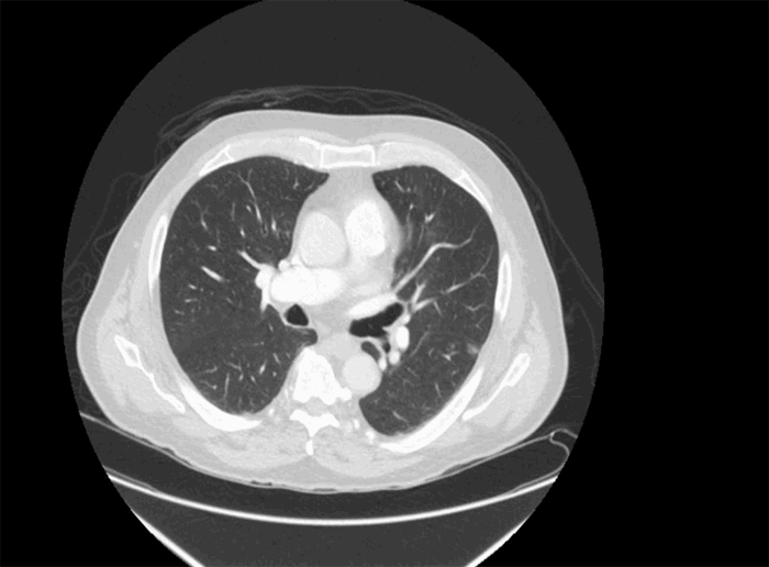
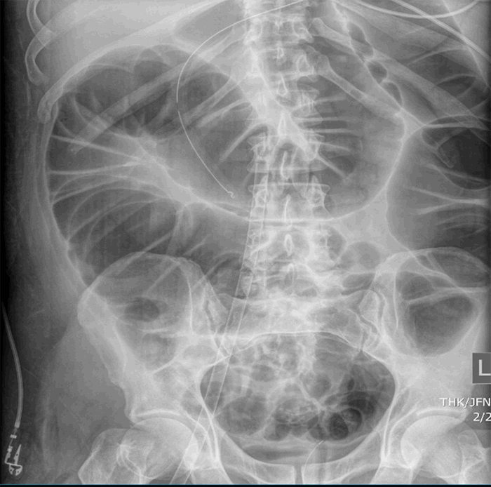
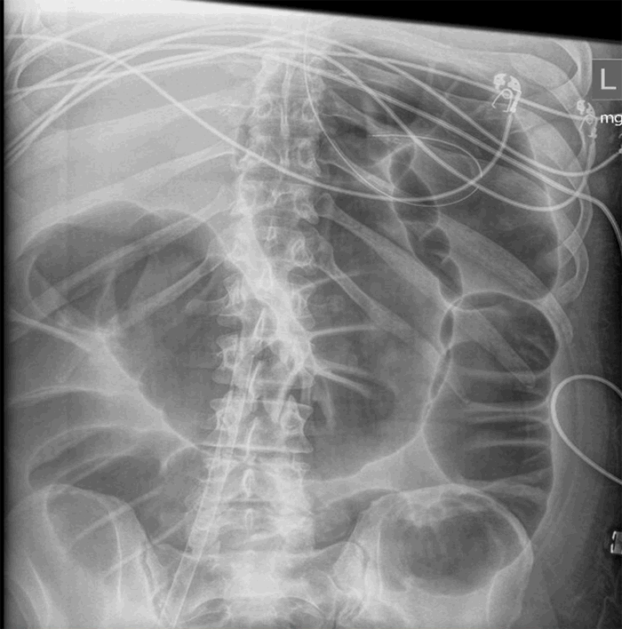
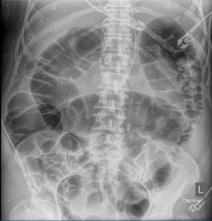
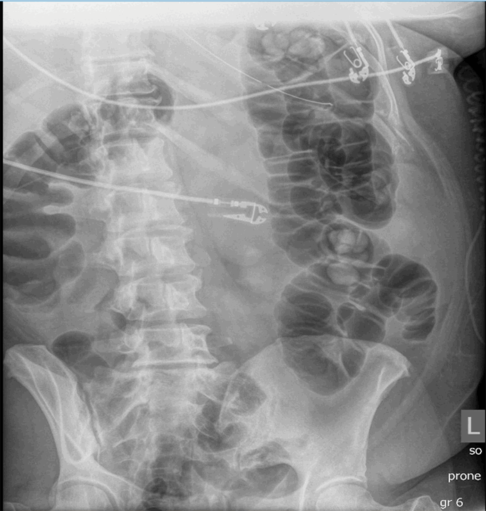
The three patients presented in this case series have several similarities as well as distinct differences (Table 1). All were COVID-19 positive with severe ARDS requiring intubation and mechanical ventilation; one required ECMO. All patients had evidence of multiple organ dysfunction syndrome, and they also all had evidence of acute colonic pseudo-obstruction, with functional dilatation of the colon but no mechanical obstruction. The patient (described in Case 1) who developed cecal ischemia had a 2 cm increase in his cecal diameter over a 24-hour period, his cecal diameter with the first KUB was 11.6 cm, his WBC doubled within 24 hours, and his lactate level continued to rise. Pneumatosis of the colon was evident on the KUB before operative intervention. Although he responded initially to nonoperative measures like the placement of a rectal tube and an aggressive bowel regimen, he went on to develop bowel ischemia. The second patient (described in Case 2) who responded to nonoperative management had a colonic diameter of 7.8 cm on the initial KUB, his colonic diameter increased by approximately 0.5 cm per day, his WBC was on the low end of normal, and his lactate was normal. The third patient (described in Case 3) responded to nonoperative management and had normal lactate as well as WBC.


Case 1

Case 2

Case 3

Age

69

49

43

Gender

Male

Male

Female

BMI (kg/m2)

25.8

26.1

33.3

WBC (uL)

6900

4000

8400

Lymphocyte%

19.3

11.0

7.8

D-dimer (ng/mL)

458

995

1361

Lactate (mg/dL), at time of diagnosis of ileus

2.9

0.9

0.8

Fibrinogen (mg/dL)

1034

811

618

Abdominal X ray

Colonic dilatation

Colonic dilatation

Colonic dilatation

Rectal tube

Yes

Yes

Yes

Bowel ischemia

Yes

No

No

Surgical intervention

Yes

No

No

ARDS

Yes

Yes

Yes

Mortality

Yes

No

No

Table 1. Summary of the major findings in these three described case reports

It is difficult to generalize based on a case series of only three patients, but clinicians taking care of COVID-positive patients should be aware of the risk of colonic paralytic ileus in this disease and its potentially devastating consequences. It remains unclear whether neostigmine would be effective in these cases as we did not use it in any of the three patients in our series, but extrapolating from Ogilvie, we would recommend its use in case of worsening of the COVID-19 paralytic ileus despite adequate bowel regimen and colonic decompression with a rectal tube. As multi-institutional data accumulates on these patients from across the United States and the world, we should investigate further the epidemiology, etiology, predictors, and outcomes of COVID-19-related paralytic colonic ileus and pseudo-obstruction.

Conclusion

In the three cases reported, paralytic ileus was present, and it progressed to bowel ischemia. Front-line physicians caring for patients with COVID-19 infection should be aware of this complication.

Lessons Learned

Paralytic ileus involving both the small and the large bowel appears to be a complication of COVID-19 infection. The potential for this to progress to frank bowel ischemia exists. Aggressive bowel regimen and close abdominal exam are both warranted in this condition.

Authors

Hwabejire JO; Saillant N; King R; Velmahos GC; Kaafarani HMA

Corresponding Author

John O. Hwabejire, MD, MPH
Division of Trauma, Emergency Surgery, and Surgical Critical Care
Department of Surgery, Massachusetts General Hospital and Harvard Medical School, Boston, MA
Phone: (617) 643-2439
E-mail: jhwabejire@partners.org

Author Affiliations

Division of Trauma, Emergency Surgery, and Surgical Critical Care, Department of Surgery, Massachusetts General Hospital
Harvard Medical School, Boston, MA

Disclosure Statement

The authors have no conflicts of interest to disclose.

References

  1. Coronavirus Disease 2019 (COVID-19), Situation Report – 91. World Health Organization https://www.who.int/docs/default-source/coronaviruse/situation-reports/20200812-covid-19-sitrep-205.pdf?sfvrsn=627c9aa8_2 Last accessed: 08/13/2020
  2. Coronavirus Disease 2019 (COVID-19), US Cases. The Centers for Disease Control and Prevention https://www.cdc.gov/coronavirus/2019-ncov/cases-updates/cases-in-us.html Last accessed: 08/13/2020
  3. El Moheb M, Naar L, Christensen MA, et al. Gastrointestinal Complications in Critically Ill Patients With and Without COVID-19 [published online ahead of print, 2020 Sep 24]. JAMA. 2020;324(18):1899-1901. doi:10.1001/jama.2020.19400
  4. Hutchinson R, Griffiths C. Acute colonic pseudo-obstruction: a pharmacological approach. Ann R Coll Surg Engl. 1992;74(5):364-367.
  5. Saunders MD, Kimmey MB. Colonic pseudo-obstruction: the dilated colon in the ICU. Semin Gastrointest Dis. 2003;14(1):20-27.
  6. Wells CI, O'Grady G, Bissett IP. Acute colonic pseudo-obstruction: A systematic review of aetiology and mechanisms. World J Gastroenterol. 2017;23(30):5634-5644. doi:10.3748/wjg.v23.i30.5634
  7. Vanek VW, Al-Salti M. Acute pseudo-obstruction of the colon (Ogilvie's syndrome). An analysis of 400 cases. Dis Colon Rectum. 1986;29(3):203-210. doi:10.1007/BF02555027
  8. Ross SW, Oommen B, Wormer BA, et al. Acute Colonic Pseudo-obstruction: Defining the Epidemiology, Treatment, and Adverse Outcomes of Ogilvie's Syndrome. Am Surg. 2016;82(2):102-111. doi:10.1177/000313481608200211
  9. Nanni G, Garbini A, Luchetti P, Nanni G, Ronconi P, Castagneto M. Ogilvie's syndrome (acute colonic pseudo-obstruction): review of the literature (October 1948 to March 1980) and report of four additional cases. Dis Colon Rectum. 1982;25(2):157-166. doi:10.1007/BF02553265
  10. Delgado-Aros S, Camilleri M. Pseudo-obstruction in the critically ill. Best Pract Res Clin Gastroenterol. 2003;17(3):427-444. doi:10.1016/s1521-6918(03)00023-4
  11. Saunders MD, Kimmey MB. Colonic pseudo-obstruction: the dilated colon in the ICU. Semin Gastrointest Dis. 2003;14(1):20-27.
  12. Maloney N, Vargas HD. Acute intestinal pseudo-obstruction (Ogilvie's syndrome). Clin Colon Rectal Surg. 2005;18(2):96-101. doi:10.1055/s-2005-870890
  13. De Giorgio R, Ricciardiello L, Naponelli V, et al. Chronic intestinal pseudo-obstruction related to viral infections. Transplant Proc. 2010;42(1):9-14. doi:10.1016/j.transproceed.2009.12.014
  14. Selgrad M, De Giorgio R, Fini L, et al. JC virus infects the enteric glia of patients with chronic idiopathic intestinal pseudo-obstruction. Gut. 2009;58(1):25-32. doi:10.1136/gut.2008.152512
  15. Sinagra E, Raimondo D, Gallo E, et al. Could JC virus be linked to chronic idiopathic intestinal pseudo-obstruction?. Clin J Gastroenterol. 2020;13(3):377-381. doi:10.1007/s12328-019-01069-4
  16. Arbour N, Day R, Newcombe J, Talbot PJ. Neuroinvasion by human respiratory coronaviruses. J Virol. 2000;74(19):8913-8921. doi:10.1128/jvi.74.19.8913-8921.2000
  17. Desforges M, Le Coupanec A, Brison E, Meessen-Pinard M, Talbot PJ. Neuroinvasive and neurotropic human respiratory coronaviruses: potential neurovirulent agents in humans. Adv Exp Med Biol. 2014;807:75-96. doi:10.1007/978-81-322-1777-0_6
  18. Xu J, Zhong S, Liu J, et al. Detection of severe acute respiratory syndrome coronavirus in the brain: potential role of the chemokine mig in pathogenesis. Clin Infect Dis. 2005;41(8):1089-1096. doi:10.1086/444461
  19. Mao L, Jin H, Wang M, et al. Neurologic Manifestations of Hospitalized Patients With Coronavirus Disease 2019 in Wuhan, China. JAMA Neurol. 2020;77(6):683-690. doi:10.1001/jamaneurol.2020.1127