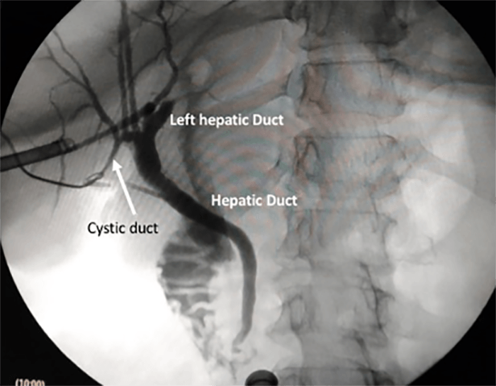
Figure 1. Intraoperative cholangiography showing cystic duct (white arrow) draining in the right hepatic duct
Discussion
Bile duct anatomic variations are described following Huang's classification, based on the insertion of right posterior bile duct.3 According to this, five distinct anatomic types have been defined. With regard to the cystic duct, the classical anatomy draining in the right lateral aspect at the middle third of the common hepatic duct is seen in 58 to 75% of patients.4 Most common anatomic variations are related to the point of insertion in the extrahepatic bile duct: medial insertion, low insertion, and parallel course.5
Abnormal drainage of the cystic duct into the right hepatic duct, as seen in our case, is extremely rare, and is estimated to occur in 0.3 to 0.4% of patients.6 Even more infrequent is the presence of a right posterolateral duct opening in the cystic duct (Huang's A5 type of biliary tree variation), an exceedingly rare anatomic variant that was also recently reported.7
At our institution, we systematically achieved Strasberg's critical view and performed intraoperative cholangiography whenever possible to assess biliary anatomy and detect unsuspected choledochal gallstones. Although anatomic variations can be sometimes detected preoperatively by different imaging modalities such as ultrasound, computed tomography, magnetic resonance cholangiography, or even endoscopic retrograde cholangiopancreatography,8 not all of these might be indicated or even available before surgery. This is not unusual when there is no suspicion of bile duct gallstones, whether by clinical findings, laboratory tests, or even imageology, just as we stated in the case of our patient. In such cases, the only way to correctly assess bile duct anatomy and its variations is to perform intraoperative cholangiography.
While some anatomic variations of the biliary tract may not be clinically relevant, it is very important to diagnose the presence of a high union of the cystic duct into the common hepatic duct, the right hepatic duct, or the intrahepatic bile ducts, since this could increase the risk of iatrogenic injuries when not properly recognized.
Conclusion
Even though some anatomic variations of the biliary tract may not be clinically relevant, it is very important to diagnose the presence of a high union of the cystic duct into the common hepatic duct, the right hepatic duct, or the intrahepatic bile ducts, since this could increase the risk of iatrogenic injuries when not properly recognized.
Lessons Learned
The case described above reinforces the importance of considering all types of anatomical variations of the biliary duct system whenever performing a cholecystectomy. If surgeons are aware of these possible findings, the risk of iatrogenic bile duct injuries will be minimized.
Authors
Javier Chinelli, MD
Clínica Quirúrgica 2 (Surgical Clinic 2)
Hospital Maciel
Montevideo, Uruguay
Emilia Moreira, MD
Clínica Quirúrgica 2 (Surgical Clinic 2)
Hospital Maciel
Montevideo, Uruguay
Juan Costa, MD
Clínica Quirúrgica 2 (Surgical Clinic 2)
Hospital Maciel
Montevideo, Uruguay
Gustavo Rodriguez, MD
Clínica Quirúrgica 2 (Surgical Clinic 2)
Hospital Maciel
Montevideo, Uruguay
Correspondence Author
Dr. Javier Chinelli
Surgeon, Clínica Quirúrgica 2
Hospital Maciel
Mercedes 1472/402
PA 11200
Montevideo, Uruguay
Phone: 598 099491516
Email: jchinelli01@gmail.com
References
- Turner MA, Fulcher, AS. The cystic duct: normal anatomy and disease processes. Radiographics 2001;21:1,3–22.
- Wu Y-H, Liu Z-S, Mrikhi R. Anatomical variations of the cystic duct: two case reports. World J Gastroenterol 2008;14:1 155–157.
- Huang TL, Cheng YF, Chen CL, Lee TY. Variants of the bile ducts: clinical application in the potential donor of living-related hepatic transplantation. Transplant Proc 1996;28:1669-1670.
- Talpur K.A.H, Laghari A.A, Yousfani S.A, Malik AM, Memon I, Khan S.A. Anatomical variations and congenital anomalies of extra hepatic biliary system encountered during laparoscopic cholecystectomy. J Pak Med Assoc 2010; 60:2 89–93.
- Sarawagi R, Sundar S, Guptar S, Raghuwanshi S. Anatomical Variations of Cystic Ducts in Magnetic Resonance Cholangiopancreatography and Clinical Implications. Radiology Research and Practice 2016;1-6 http://dx.doi.org/10.1155/2016/3021484
- Carbajo MA, Martin del Omo JC, Blanco JI. Congenital malformations of the gallbladder and cystic duct diagnosed by laparoscopy: high surgical risk. J Soc Laparoendo 1999;3:4 319–321.
- Hernández R, Pontillo, M, Chinelli J, Rodriguez G. Colangiography: rare anatomical variation of the bile duct. Cir Esp 2018. https://doi.org/ 10.1016/j.ciresp.2018.05.006
- Taourel P, Bret PM, Reinhold C, Barkun AN, Atri M. Anatomic variants of the biliary tree: diagnosis with MR cholangiopancreatography. Radiology 1996;199:521–7.